We use cookies to enhance the usability of our website. If you continue, we'll assume that you are happy to receive all cookies. More information. Don't show this again.
Fields »
 

Field

Term
Gene name

Class

Subclass

Chromosome

External id

In atlas

Reliability

Reliability

Reliability

Validation

Method

Validation

Method

Validation

Method

Validation

Location

Searches

Tissue

Cell type

Expression

Tissue

Specificity

Cell line

Specificity

Tissue

Detectable

Cell line

Detectable

Category

Score

Score

Score

Antibodies

Column


ABOUT
HELP
BLOG
  • THE HUMAN PROTEOME
  • THE HUMAN TISSUES
  • THE HUMAN CELL
  • PROTEIN CLASSES
  • PROTEIN EVIDENCE
    • NEWS
    • BLOG
    • EVENTS
    • PRESS ROOM
    • LEARN
    • DICTIONARY
    • METHODS
    • CELL LINES
  • THE PROJECT
  • INTRODUCTION
  • ORGANIZATION
  • PUBLICATIONS
  • PUBLICATION DATA
  • ANTIBODY SUBMISSION
  • ANTIBODY AVAILABILITY
  • LINKS
  • CONTACT
  • TECHNICAL DATA
  • ANTIBODY VALIDATION
  • ASSAYS & ANNOTATION
  • DISCLAIMER
  • DOWNLOADABLE DATA
  • HELP & FAQ
  • LICENCE & CITATION
  • PRIVACY STATEMENT
  • RELEASE HISTORY
PSMB1
  • premium

    TISSUE

  • premium

    CELL

  • CANCER

CANCER ATLAS

GENE/PROTEIN

Antibody validation

Dictionary

Level of antibody staining/expression


High
Medium
Low
Not detected

CANCER ATLASi

The cancer tissue page shows antibody staining in 20 different cancers. The assay and annotation is described here.

This page starts with information about the protein evidence and, when applicable, also protein class. Below the summary, a selection of four standard cancer tissue samples is displayed as representative of the overall staining pattern. From left: colorectal cancer, breast cancer, prostate cancer and lung cancer. An additional 5th image can be added as a complement.

For histological reference, visit the histological dictionary.

Gene description

Proteasome subunit beta 1

Protein class

Enzymes, FDA approved drug targets, Plasma proteins, Predicted intracellular proteins

Protein evidence

Evidence at protein level
Colorectal cancer
Breast cancer
Prostate cancer
Lung cancer
Liver cancer

STAINING SUMMARYi

The cancer tissue summary page shows antibody staining in 20 different cancers. The assay and annotation is described here.

For each cancer, the fraction of samples with antibody staining/protein expression level high, medium, low, or not detected are provided by the blue-scale color-coding (as described by the color-coding scale in the box to the right). The length of the bar represents the number of patient samples analyzed (max=12 patients). The images and annotations can be accessed by clicking on the cancer name or protein expression bar. If more than one antibody is analyzed, the tabs at the top of the staining summary section can be used to toggle between the different antibodies. The tooltip function displays additional data for the features in the staining summary view.

Next to the cancer staining data, the protein expression data of normal tissues or specific cell types corresponding to each cancer are shown and protein expression levels are indicated by the blue-scale color coding.

At the bottom of the page, a summary of the overall protein expression pattern across the analyzed cancer tissues as well as information about literature conformity are presented.

HPA029635
HPA029637
CAB033911

Tissue

Cancer staining

Protein
expression of
normal tissue

Breast cancer
 
Carcinoid
 
Cervical cancer
 
 
Colorectal cancer
 
 
Endometrial cancer
 
 
Glioma
 
Head and neck cancer
 
 
Liver cancer
 
 
Lung cancer
 
 
Lymphoma


Tissue

Cancer staining

Protein
expression of
normal tissue

Melanoma
 
Ovarian cancer
 
Pancreatic cancer
 
Prostate cancer
 
Renal cancer
 
Skin cancer
 
Stomach cancer
 
 
Testis cancer
 
Thyroid cancer
 
Urothelial cancer
 

Staining summary

Most cancer cells showed weak to moderate positivity. Several cases of carcinoids, malignant melanoma, liver and colorectal cancers displayed strong cytoplasmic and nuclear immunoreactivity.Remaining cancer tissues were weakly stained or negative.

Tissue

Cancer staining

Protein
expression of
normal tissue

Breast cancer
 
Carcinoid
 
Cervical cancer
 
 
Colorectal cancer
 
 
Endometrial cancer
 
 
Glioma
 
Head and neck cancer
 
 
Liver cancer
 
 
Lung cancer
 
 
Lymphoma


Tissue

Cancer staining

Protein
expression of
normal tissue

Melanoma
 
Ovarian cancer
 
Pancreatic cancer
 
Prostate cancer
 
Renal cancer
 
Skin cancer
 
Stomach cancer
 
 
Testis cancer
 
Thyroid cancer
 
Urothelial cancer
 

Staining summary

Most cancer cells displayed moderate to strong cytoplasmic and nuclear immunoreactivity. Few cases of carcinoid and head & neck cancers were negative.

Tissue

Cancer staining

Protein
expression of
normal tissue

Breast cancer
 
Carcinoid
 
Cervical cancer
 
 
Colorectal cancer
 
 
Endometrial cancer
 
 
Glioma
 
Head and neck cancer
 
 
Liver cancer
 
 
Lung cancer
 
 
Lymphoma


Tissue

Cancer staining

Protein
expression of
normal tissue

Melanoma
 
Ovarian cancer
 
Pancreatic cancer
 
Prostate cancer
 
Renal cancer
 
Skin cancer
 
Stomach cancer
 
 
Testis cancer
 
Thyroid cancer
 
Urothelial cancer
 

Staining summary

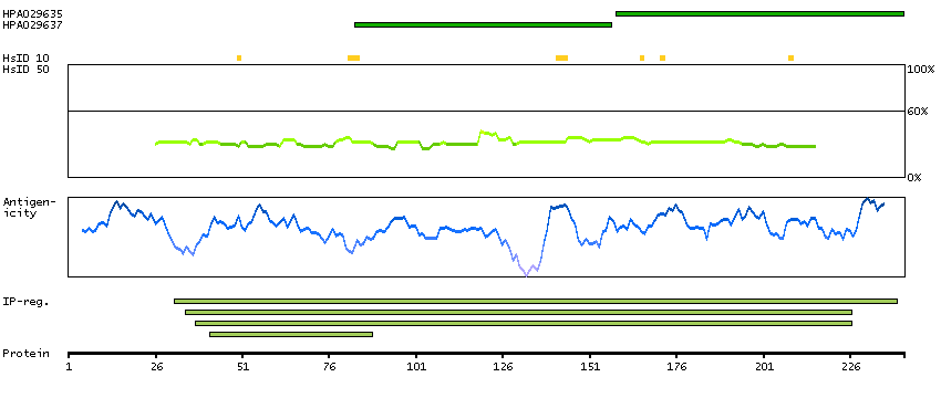
Most cancers showed moderate to strong cytoplasmic and nuclear immunoreactivity. Basal cell carcinomas, several ovarian and stomach cancers were weakly stained.

GENE INFORMATIONi

Gene information from Ensembl and Entrez, as well as links to available gene identifiers are displayed here. Information was retrieved from Ensembl if not indicated otherwise.

Gene name

PSMB1 (HGNC Symbol)

Synonyms

HC5, PMSB1

Description

Proteasome subunit beta 1 (HGNC Symbol)

Entrez gene summary

The proteasome is a multicatalytic proteinase complex with a highly ordered ring-shaped 20S core structure. The core structure is composed of 4 rings of 28 non-identical subunits; 2 rings are composed of 7 alpha subunits and 2 rings are composed of 7 beta subunits. Proteasomes are distributed throughout eukaryotic cells at a high concentration and cleave peptides in an ATP/ubiquitin-dependent process in a non-lysosomal pathway. An essential function of a modified proteasome, the immunoproteasome, is the processing of class I MHC peptides. This gene encodes a member of the proteasome B-type family, also known as the T1B family, that is a 20S core beta subunit. This gene is tightly linked to the TBP (TATA-binding protein) gene in human and in mouse, and is transcribed in the opposite orientation in both species. [provided by RefSeq, Jul 2008]

Chromosome

6

Cytoband

q27

Chromosome location (bp)

170535117 - 170553341

Protein evidence

Evidence at protein level (all genes)

Ensembl

ENSG00000008018 (version 83.38)

Entrez gene

5689

UniProt

P20618 (UniProt - Evidence at protein level)

neXtProt

NX_P20618

Antibodypedia

PSMB1 antibodies

PROTEIN BROWSERi

The protein browser displays the antigen location on the target protein(s) and the features of the target protein. The tabs at the top of the protein view section can be used to switch between the different splice variants to which an antigen has been mapped.

At the top of the view, the position of the antigen (identified by the corresponding HPA identifier) is shown as a green bar. A yellow triangle on the bar indicates a <100% sequence identity to the protein target.

Under the antigens, the maximum percent sequence identity of the protein to all other proteins from other human genes is displayed, using a sliding window of 10 aa residues (HsID 10) or 50 aa residues (HsID 50) (read more).

If a signal peptide is predicted by a majority of the signal peptide predictors SPOCTOPUS, SignalP 4.0, and Phobius (turquoise) and/or transmembrane regions (orange) are predicted by MDM, these are displayed.

Low complexity regions are shown in yellow and InterPro regions in green. Common (purple) and unique (grey) regions between different splice variants of the gene are also displayed (read more), and at the bottom of the protein view is the protein scale.

PSMB1-001

PROTEIN INFORMATIONi

The protein information section displays alternative protein-coding transcripts (splice variants) encoded by this gene according to the Ensembl database.

The ENSP identifier links to the Ensembl website protein summary, while the ENST identifier links to the Ensembl website transcript summary for the selected splice variant. The data in the UniProt column can be expanded to show links to all matching UniProt identifiers for this protein.

The protein classes assigned to this protein are shown if expanding the data in the protein class column. Parent protein classes are in bold font and subclasses are listed under the parent class.

The Gene Ontology terms assigned to this protein are listed if expanding the Gene ontology column. The length of the protein (amino acid residues according to Ensembl), molecular mass (kDalton), predicted signal peptide (according to a majority of the signal peptide predictors SPOCTOPUS, SignalP 4.0, and Phobius) and the number of predicted transmembrane region(s) (according to MDM) are also reported.

Splice variant

UniProt

Protein class

Gene ontology

Length & mass

Signal peptide
(predicted)

Transmembrane regions
(predicted)

PSMB1-001
ENSP00000262193
ENST00000262193
P20618 [Direct mapping]
Proteasome subunit beta type-1
Show all
Enzymes
   ENZYME proteins
   Hydrolases
   Peptidases
   Threonine-type peptidases
Predicted intracellular proteins
Plasma proteins
FDA approved drug targets
   Small molecule drugs
Protein evidence (Kim et al 2014)
Protein evidence (Ezkurdia et al 2014)
Show all
GO:0000082 [G1/S transition of mitotic cell cycle]
GO:0000165 [MAPK cascade]
GO:0000186 [activation of MAPKK activity]
GO:0000209 [protein polyubiquitination]
GO:0000278 [mitotic cell cycle]
GO:0000502 [proteasome complex]
GO:0002223 [stimulatory C-type lectin receptor signaling pathway]
GO:0002474 [antigen processing and presentation of peptide antigen via MHC class I]
GO:0002479 [antigen processing and presentation of exogenous peptide antigen via MHC class I, TAP-dependent]
GO:0004175 [endopeptidase activity]
GO:0004298 [threonine-type endopeptidase activity]
GO:0005515 [protein binding]
GO:0005634 [nucleus]
GO:0005654 [nucleoplasm]
GO:0005737 [cytoplasm]
GO:0005829 [cytosol]
GO:0005839 [proteasome core complex]
GO:0006521 [regulation of cellular amino acid metabolic process]
GO:0006595 [polyamine metabolic process]
GO:0006915 [apoptotic process]
GO:0006977 [DNA damage response, signal transduction by p53 class mediator resulting in cell cycle arrest]
GO:0007173 [epidermal growth factor receptor signaling pathway]
GO:0007264 [small GTPase mediated signal transduction]
GO:0007265 [Ras protein signal transduction]
GO:0007411 [axon guidance]
GO:0008286 [insulin receptor signaling pathway]
GO:0008543 [fibroblast growth factor receptor signaling pathway]
GO:0010467 [gene expression]
GO:0010499 [proteasomal ubiquitin-independent protein catabolic process]
GO:0012501 [programmed cell death]
GO:0016032 [viral process]
GO:0031145 [anaphase-promoting complex-dependent proteasomal ubiquitin-dependent protein catabolic process]
GO:0033209 [tumor necrosis factor-mediated signaling pathway]
GO:0034641 [cellular nitrogen compound metabolic process]
GO:0038061 [NIK/NF-kappaB signaling]
GO:0038095 [Fc-epsilon receptor signaling pathway]
GO:0042590 [antigen processing and presentation of exogenous peptide antigen via MHC class I]
GO:0042981 [regulation of apoptotic process]
GO:0043066 [negative regulation of apoptotic process]
GO:0043161 [proteasome-mediated ubiquitin-dependent protein catabolic process]
GO:0043488 [regulation of mRNA stability]
GO:0044281 [small molecule metabolic process]
GO:0045087 [innate immune response]
GO:0048010 [vascular endothelial growth factor receptor signaling pathway]
GO:0048011 [neurotrophin TRK receptor signaling pathway]
GO:0050852 [T cell receptor signaling pathway]
GO:0051436 [negative regulation of ubiquitin-protein ligase activity involved in mitotic cell cycle]
GO:0051437 [positive regulation of ubiquitin-protein ligase activity involved in regulation of mitotic cell cycle transition]
GO:0051439 [regulation of ubiquitin-protein ligase activity involved in mitotic cell cycle]
GO:0051603 [proteolysis involved in cellular protein catabolic process]
GO:0070062 [extracellular exosome]
GO:0090090 [negative regulation of canonical Wnt signaling pathway]
GO:0090263 [positive regulation of canonical Wnt signaling pathway]
Show all
241 aa
26.5 kDa
No 0

Contact

  • BLOG
  • PRESS ROOM
  • contact@proteinatlas.org

The Project

  • INTRODUCTION
  • ORGANIZATION
  • PUBLICATIONS
  • DOWNLOADABLE DATA

The Human Protein Atlas

  • ANTIBODY AVAILABILITY
  • LICENCE & CITATION
  • HELP & FAQ
The Human Protein Atlas project is funded
by the Knut & Alice Wallenberg foundation.