We use cookies to enhance the usability of our website. If you continue, we'll assume that you are happy to receive all cookies. More information. Don't show this again.
Fields »
 

Field

Term
Gene name

Class

Subclass

Chromosome

External id

In atlas

Reliability

Reliability

Reliability

Validation

Method

Validation

Method

Validation

Method

Validation

Location

Searches

Tissue

Cell type

Expression

Tissue

Specificity

Cell line

Specificity

Tissue

Detectable

Cell line

Detectable

Category

Score

Score

Score

Antibodies

Column


ABOUT
HELP
BLOG
  • THE HUMAN PROTEOME
  • THE HUMAN TISSUES
  • THE HUMAN CELL
  • PROTEIN CLASSES
  • PROTEIN EVIDENCE
    • NEWS
    • BLOG
    • EVENTS
    • PRESS ROOM
    • LEARN
    • DICTIONARY
    • METHODS
    • CELL LINES
  • THE PROJECT
  • INTRODUCTION
  • ORGANIZATION
  • PUBLICATIONS
  • PUBLICATION DATA
  • ANTIBODY SUBMISSION
  • ANTIBODY AVAILABILITY
  • LINKS
  • CONTACT
  • TECHNICAL DATA
  • ANTIBODY VALIDATION
  • ASSAYS & ANNOTATION
  • DISCLAIMER
  • DOWNLOADABLE DATA
  • HELP & FAQ
  • LICENCE & CITATION
  • PRIVACY STATEMENT
  • RELEASE HISTORY
ADH1C
  • premium

    TISSUE

  • CELL

  • CANCER

CANCER ATLAS

GENE/PROTEIN

Antibody validation

Dictionary

Level of antibody staining/expression


High
Medium
Low
Not detected

CANCER ATLASi

The cancer tissue page shows antibody staining in 20 different cancers. The assay and annotation is described here.

This page starts with information about the protein evidence and, when applicable, also protein class. Below the summary, a selection of four standard cancer tissue samples is displayed as representative of the overall staining pattern. From left: colorectal cancer, breast cancer, prostate cancer and lung cancer. An additional 5th image can be added as a complement.

For histological reference, visit the histological dictionary.

Gene description

Alcohol dehydrogenase 1C (class I), gamma polypeptide

Protein class

Enzymes, FDA approved drug targets, Plasma proteins, Predicted intracellular proteins

Protein evidence

Evidence at protein level
Colorectal cancer
Breast cancer
Prostate cancer
Lung cancer

STAINING SUMMARYi

The cancer tissue summary page shows antibody staining in 20 different cancers. The assay and annotation is described here.

For each cancer, the fraction of samples with antibody staining/protein expression level high, medium, low, or not detected are provided by the blue-scale color-coding (as described by the color-coding scale in the box to the right). The length of the bar represents the number of patient samples analyzed (max=12 patients). The images and annotations can be accessed by clicking on the cancer name or protein expression bar. If more than one antibody is analyzed, the tabs at the top of the staining summary section can be used to toggle between the different antibodies. The tooltip function displays additional data for the features in the staining summary view.

Next to the cancer staining data, the protein expression data of normal tissues or specific cell types corresponding to each cancer are shown and protein expression levels are indicated by the blue-scale color coding.

At the bottom of the page, a summary of the overall protein expression pattern across the analyzed cancer tissues as well as information about literature conformity are presented.

HPA047814
HPA060902

Tissue

Cancer staining

Protein
expression of
normal tissue

Breast cancer
 
Carcinoid
 
Cervical cancer
 
 
Colorectal cancer
 
 
Endometrial cancer
 
 
Glioma
 
Head and neck cancer
 
 
Liver cancer
 
 
Lung cancer
 
 
Lymphoma


Tissue

Cancer staining

Protein
expression of
normal tissue

Melanoma
 
Ovarian cancer
 
Pancreatic cancer
 
Prostate cancer
 
Renal cancer
 
Skin cancer
 
Stomach cancer
 
 
Testis cancer
 
Thyroid cancer
 
Urothelial cancer
 

Staining summary

Moderate to strong cytoplasmic and nuclear staining was observed in liver cancers. Several cases of lymphomas along with a few ovarian, colorectal, breast and prostate cancers showed weak to moderate staining. Remaining cancer tissues were mainly negative.

Tissue

Cancer staining

Protein
expression of
normal tissue

Breast cancer
 
Carcinoid
 
Cervical cancer
 
 
Colorectal cancer
 
 
Endometrial cancer
 
 
Glioma
 
Head and neck cancer
 
 
Liver cancer
 
 
Lung cancer
 
 
Lymphoma


Tissue

Cancer staining

Protein
expression of
normal tissue

Melanoma
 
Ovarian cancer
 
Pancreatic cancer
 
Prostate cancer
 
Renal cancer
 
Skin cancer
 
Stomach cancer
 
 
Testis cancer
 
Thyroid cancer
 
Urothelial cancer
 

Staining summary

Few hepatocellular carcinomas displayed moderate cytoplasmic staining. Remaining cancer tissues were negative.

GENE INFORMATIONi

Gene information from Ensembl and Entrez, as well as links to available gene identifiers are displayed here. Information was retrieved from Ensembl if not indicated otherwise.

Gene name

ADH1C (HGNC Symbol)

Synonyms

ADH3

Description

Alcohol dehydrogenase 1C (class I), gamma polypeptide (HGNC Symbol)

Entrez gene summary

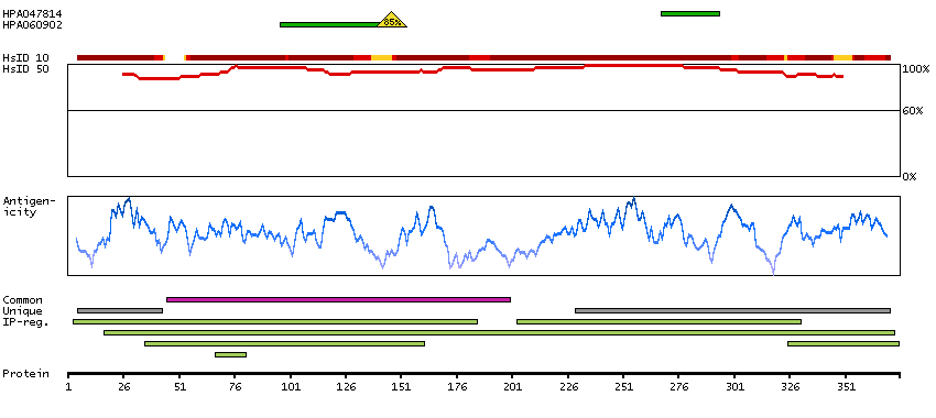
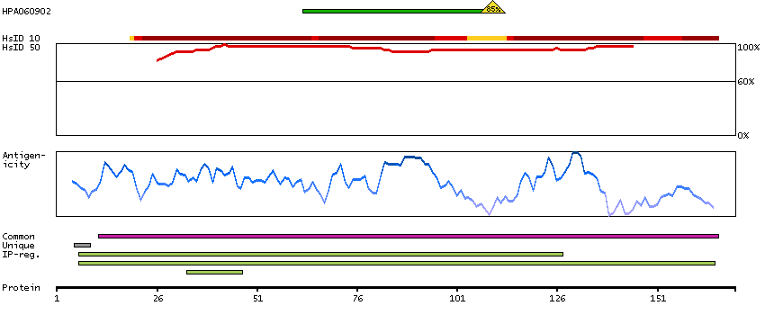
This gene encodes class I alcohol dehydrogenase, gamma subunit, which is a member of the alcohol dehydrogenase family. Members of this enzyme family metabolize a wide variety of substrates, including ethanol, retinol, other aliphatic alcohols, hydroxysteroids, and lipid peroxidation products. Class I alcohol dehydrogenase, consisting of several homo- and heterodimers of alpha, beta, and gamma subunits, exhibits high activity for ethanol oxidation and plays a major role in ethanol catabolism. Three genes encoding alpha, beta and gamma subunits are tandemly organized in a genomic segment as a gene cluster. [provided by RefSeq, Jul 2008]

Chromosome

4

Cytoband

q23

Chromosome location (bp)

99336492 - 99353027

Protein evidence

Evidence at protein level (all genes)

Ensembl

ENSG00000248144 (version 83.38)

Entrez gene

126

UniProt

P00326 (UniProt - Evidence at protein level)

neXtProt

NX_P00326

Antibodypedia

ADH1C antibodies

PROTEIN BROWSERi

The protein browser displays the antigen location on the target protein(s) and the features of the target protein. The tabs at the top of the protein view section can be used to switch between the different splice variants to which an antigen has been mapped.

At the top of the view, the position of the antigen (identified by the corresponding HPA identifier) is shown as a green bar. A yellow triangle on the bar indicates a <100% sequence identity to the protein target.

Under the antigens, the maximum percent sequence identity of the protein to all other proteins from other human genes is displayed, using a sliding window of 10 aa residues (HsID 10) or 50 aa residues (HsID 50) (read more).

If a signal peptide is predicted by a majority of the signal peptide predictors SPOCTOPUS, SignalP 4.0, and Phobius (turquoise) and/or transmembrane regions (orange) are predicted by MDM, these are displayed.

Low complexity regions are shown in yellow and InterPro regions in green. Common (purple) and unique (grey) regions between different splice variants of the gene are also displayed (read more), and at the bottom of the protein view is the protein scale.

ADH1C-001
ADH1C-002
ADH1C-004

PROTEIN INFORMATIONi

The protein information section displays alternative protein-coding transcripts (splice variants) encoded by this gene according to the Ensembl database.

The ENSP identifier links to the Ensembl website protein summary, while the ENST identifier links to the Ensembl website transcript summary for the selected splice variant. The data in the UniProt column can be expanded to show links to all matching UniProt identifiers for this protein.

The protein classes assigned to this protein are shown if expanding the data in the protein class column. Parent protein classes are in bold font and subclasses are listed under the parent class.

The Gene Ontology terms assigned to this protein are listed if expanding the Gene ontology column. The length of the protein (amino acid residues according to Ensembl), molecular mass (kDalton), predicted signal peptide (according to a majority of the signal peptide predictors SPOCTOPUS, SignalP 4.0, and Phobius) and the number of predicted transmembrane region(s) (according to MDM) are also reported.

Splice variant

UniProt

Protein class

Gene ontology

Length & mass

Signal peptide
(predicted)

Transmembrane regions
(predicted)

ADH1C-001
ENSP00000426083
ENST00000515683
P00326 [Direct mapping]
Alcohol dehydrogenase 1C
Show all
Enzymes
   ENZYME proteins
   Oxidoreductases
   SPOCTOPUS predicted membrane proteins
Predicted intracellular proteins
Plasma proteins
FDA approved drug targets
   Small molecule drugs
Protein evidence (Kim et al 2014)
Show all
GO:0004022 [alcohol dehydrogenase (NAD) activity]
GO:0005829 [cytosol]
GO:0006069 [ethanol oxidation]
GO:0006805 [xenobiotic metabolic process]
GO:0008270 [zinc ion binding]
GO:0016491 [oxidoreductase activity]
GO:0044281 [small molecule metabolic process]
GO:0055114 [oxidation-reduction process]
Show all
375 aa
39.9 kDa
No 0
ADH1C-002
ENSP00000478545
ENST00000511397
A0A087WUC4 [Direct mapping]
Alcohol dehydrogenase 1C
Show all
   SPOCTOPUS predicted membrane proteins
Predicted intracellular proteins
Show all
GO:0008270 [zinc ion binding]
GO:0016491 [oxidoreductase activity]
GO:0055114 [oxidation-reduction process]
Show all
170 aa
17.5 kDa
No 0
ADH1C-004
ENSP00000478439
ENST00000510055
A0A087WU81 [Direct mapping]
Alcohol dehydrogenase 1C
Show all
   SPOCTOPUS predicted membrane proteins
Predicted intracellular proteins
Show all
GO:0008270 [zinc ion binding]
GO:0016491 [oxidoreductase activity]
GO:0055114 [oxidation-reduction process]
Show all
192 aa
19.8 kDa
No 0

Contact

  • BLOG
  • PRESS ROOM
  • contact@proteinatlas.org

The Project

  • INTRODUCTION
  • ORGANIZATION
  • PUBLICATIONS
  • DOWNLOADABLE DATA

The Human Protein Atlas

  • ANTIBODY AVAILABILITY
  • LICENCE & CITATION
  • HELP & FAQ
The Human Protein Atlas project is funded
by the Knut & Alice Wallenberg foundation.