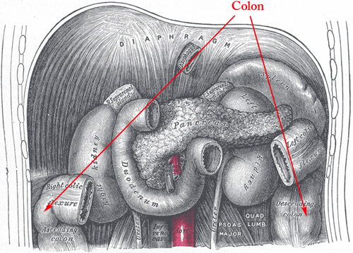
Colon
The colon is divided into four parts, the ascending, transverse, descending and sigmoid colon and is on average 1,5 meters long. Its main function is reassertion of fluid, electrolytes, and vitamins.
Since the large intestine has no villi or plica circularis the mucosa is smooth. Simple tubular intestinal glands (crypts of Lieberkuhn) extend through the entire thickness of the mucosa. The surface columnar epithelium and the cells lining the crypts are enterocytes, with an oval basal nucleus and apical brush border, the microscopic representation of microvilli. There are also numerous mucous secreting goblet cells recognized by their content of a large mucous globule. The lamina propria with connective tissue and inflammatory cells surround the crypts. A thin smooth muscular layer, the lamina muscularis mucosae marks the border between the mucosa and submucosa.
The submucosa consists of loose connective tissue with vessels and nerves. Some solitary lymph follicles are also seen.
The muscular layer (muscularis externa) consists of an inner circular smooth muscle layer, the outer longitudinal muscle layer is not continuous as in the rest of the gastrointestinal tract. It is divided into three thickened muscular bands, called taenia coli.
General histology of gastrointestinal tract (GI-tract)
The gastrointestinal canal consists of the esophagus, stomach, duodenum, jejunum, ileum, colon, rectum and anal canal. It is best viewed as a long tube passing from the oral to the anal opening. It supplies the body with water, electrolytes and nutrients from the food we eat. Our main sources of food are carbohydrates, proteins and fats, which in general cannot be absorbed in the form they are ingested. First they have to be broken down into small enough compounds. The process of digestion and absorption is carried out in a stepwise fashion as the food passes down the different parts of the gastrointestinal tract.
The general structure of all parts of the GI-tract is
1) tunica serosa /adventitia - Loose connective tissue with elastic and collagen fibers, nerves and vessels, covered by a single layer of flat mesothelial cells. Where there is no mesothelial cover the outermost layer is called adventitia.
2) tela subserosa - thin layer of loose connective tissue separating the serosa and muscle layer.
3) tunica muscularis - which for most parts is composed of an inner circular and outer longitudinal smooth muscle layer. Between the muscle fibers the myenteric plexus of Auerbach can be identified.
4) tela submucosa - a thick layer of loose connective tissue with numerous of blood and lymphatic vessels. Here is where the ganglion cells of the submucosal plexus of Meissner might be seen.
5) tunica mucosa - the innermost layer that comes in contact with the gastrointestinal content. It has secretory and absorptive function. The mucosa consists of the innermost epithelium that forms surface cells and glands, embedded in the lamina propria containing mainly of loose connective tissue with small blood vessels and immune cells. A thin layer of smooth muscle, lamina muscularis mucosae, demarcates the division of the mucosa and submucosa.
Cancer: Colorectal cancer