Kidney
Kidney
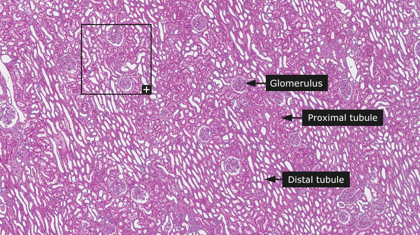
The kidneys form the first part of the urinary system and their principle function is to maintain homeostasis by the regulation of electrolytes and the acid-base balance. Kidney function is vital for regulating blood pressure and the kidneys are also a source for several important hormones such as erythropoietin, which regulates the production of red blood cells. Histologically, the renal parenchyma consists of four parts: glomeruli, tubules, interstitium and blood vessels. Glomeruli are complex vascular structures composed of a tuft of capillaries comprised of specialized endothelial, epithelial and mesangial cells arranged around a relatively thick basement membrane. The glomerulus arises from the afferent arteriole to form lobules then rejoin the vascular pole to drain into the efferent arteriole. Normally the lobules are poorly defined but highlighted in some disease processes. The tuft of capillaries lies within the lumen of the expanded proximal end of the nephron, or Bowman's space, which is lined on its parietal aspect by a layer of attenuated epithelial cells overlying a thick basement membrane. Together the epithelial cells and basement membrane comprise the Bowman's capsule. The function of the glomerulus is filtration of the blood that leads to the formation of urine.
A complex tubular system begins at the urinary pole (where urine is first formed in the Bowman's space) that extends to the renal papilla. The system comprises the proximal tubule, the loop of Henle, distal tubule and collecting duct. The proximal tubule consists of convoluted and straight portions, lined by tall columnar cells with abundant, acidophilic cytoplasm rich in structures for active fluid transport. The loop of Henle has thin descending and thick ascending portions lined by cuboidal and columnar cells. The distal tubule is narrower and shorter than the proximal tubule and lined by low cuboidal cells that do not display the deeply acidophilic, granular cytoplasm characteristic of the proximal tubule. Cuboidal cells with pale acidophilic cytoplasm and central nuclei line the collecting ducts.
The interstitium is more easily conceptualized as a space than a structure; it is visualized only when abnormal. The interstitium contains specialized interstitial cells and connective tissue elements. The larger renal blood vessels are structurally similar to those in other body sites.
Cancer: Renal cancer
|