Spleen
Spleen
The spleen is located inside the abdominal cavity on the upper left side. A normal spleen weighs about 150 grams and is about 10 cm in length. It has a rich blood supply and filters high volumes of blood. In the process it is able to detect antigens and select damaged and old red blood cells and platelets for destruction.
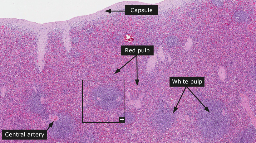
The spleen consists of red pulp and white pulp within a meshwork of reticular fibers enclosed by a dense connective tissue capsule. Blood vessels, lymph vessels and nerves enter and leave the spleen through the hilum, an opening in the capsule on the medial side. From the capsule connective tissue trabeculae extend into the organ. The trabeculae form a path for branches of the splenic artery, from which smaller arteries (central arteries) branch off to enter the pulp.
The main constituents of the spleen, the white and red pulp, form two functionally and morphologically different units. The white pulp consists of lymphatic tissue and monitors the incoming blood for harmful substances. The densely packed heterochromatin of the lymphocyte nuclei is responsible for the dark blue staining observed when sections from normal spleen are stained with HE. Aggregations of lymphocytes envelop the central arteries in a periarterial lymphatic sheath (PALS). These are mainly T-cells. At places the white pulp expands into greater spherical aggregations to form splenic nodules containing a light germinal center, consisting of proliferating B-cells, surrounding the B-cells is a darker stained mantel zone that marks the border to the red pulp. The splenic nodules have an appearance similar to lymph follicles, except for the presence of a central artery.
The red pulp filters blood to find damaged and old red blood cells and platelets. Cells that are selected for breakdown are phagocytized by splenic macrophages. The abundance of red blood cells, erythrocytes, in red pulp is accountable for the red, eosin staining observed when stained with HE. The red pulp consists of splenic cords and splenic sinuses. A meshwork of reticular cells and fibers, together with dendritic cells, macrophages, a relatively smaller amount of lymphatic cells and a great number of red blood cells constitute the splenic cords. Branches of the central arteries penetrate into the red pulp where they further branch into smaller macrophage sheathed capillaries. Within the splenic cords, the red blood cells are exposed to the macrophages and can be selected for breakdown.
|