Cerebellum

Cerebellum
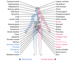
Normal brain tissue is represented by four different regions: Cerebellum, Cerebral cortex, Hippocampus and Lateral ventricle wall.
The nervous system represents the major communication network and consists of the central nervous system (CNS) and peripheral nervous system (PNS). The intracranial cerebrum and cerebellum together with the spinal cord constitutes the CNS. The brain is covered by layers of membranes, the meninges, and submerged in cerebrospinal fluid, which also fills the intracerebral ventricles. The brain can grossly be divided into different neuroanatomical functional regions such as the frontal, parietal, temporal, occipital lobes and central gray matter structures. Anatomically and histologically the brain can further be stratified into the cerebral cortex representing the outermost gray matter overlying white matter and the innermost deep gray matter components. The hippocampus, containing the neuron rich dentate fascia, is closely associated with the cerebral cortex, and is located in the medial temporal lobe. The cerebral cortex incorporates neurons (nerve cells) and glial cells (supportive cells), whereas the white matter incorporates primarily glial cells and myelinated axons from neurons.
The brain parenchyma is composed of neurons embedded in a framework of glial cells (astrocytes and oligodendrocytes) as well as microglia and blood vessels. The ependymal cell is also a specialized glial cell that lines the ventricles, including the lateral ventricles, and is closely related to the cellular component of the cerebrospinal fluid producing choroid plexus. In addition to the cell bodies that can be defined in the microscope, cell processes from neurons and glial cells form a synaptically rich "background substance" denoted as the neuropil. The cerebellum, important for coordination, appears as a highly ordered tissue with distinct layers including the cell dense granular layer and the fiber rich but sparsely cell populated molecular layer, between which the large Purkinje cells (specialized neuronal cells) are located.
The neurons are a morphologically and functionally heterogeneous family of cells that can transmit information through chemical and electrical signaling. Neurons vary in size from the small round cells that populate the internal granular layer of the cerebellum to the large pyramidal neurons of the primary motor cortex and the Purkinje cells of the cerebellum. Astrocytes represent the major glial cell type in the brain and are characterized by their cellular cytoplasmic processes reaching both synapses and capillary walls. The astrocyte is a star shaped cell involved in the maintenance of the microenvironment surrounding neurons and also important for the blood-brain barrier function. Oligodendrocytes are the main producer of myelin and are characterized by their small, rounded and lymphocyte like nuclei.
|