Duodenum
The most proximal and widest part of the small intestine is the duodenum. It starts at the pylorus of the stomach, ends at the duodenojejunal junction and measures about 25 cm long. It receives partly digested food (chyme) from the stomach and bile and pancreatic fluids from the pancreaticobiliary duct. After entering the duodenum the acidic contents from the stomach is neutralized by secretion from the intestine and pancreas. Enzymes secreted from the pancreas starts the degradation of lipids, carbohydrates and proteins to enable absorption.
As in all of the small intestine, the mucosa forms fingerlike projections called villi.
that extend into the intestinal lumen. These are epithelial folds lined by two types of cells,
enterocytes and
goblet cells. Enterocytes are
simple columnar cells with basal elongated nuclei and an apical brush border. The brush border is the microscopic representation of small protrusions of the cell membrane,
microvilli, which greatly increase the surface area of the cell enhancing absorptive capacity. The other cell type is mucus secreting goblet cells that can be recognized by the presence of an apical mucous cup. The core of the villus is part of the
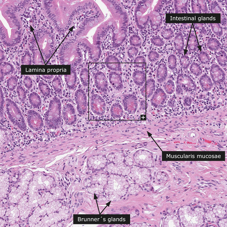
lamina propria. The most numerous cells in the lamina propria are immune cells, most of which are lymphocytes. Because villi are the site of absorption of nutrition they have a rich blood supply, each villus is supplied by central arterioles and drained by central venules and a central lymph vessel.
Underlying the villi are the intestinal glands, also called the crypts of Lieberkühn. These glands are lined with numerous relatively undifferentiated columnar cells that usually undergo two rounds of mitosis before differentiating into either absorptive cells or goblet cells. Enterocytes, goblet cells, paneth cells that secrete antibacterial enzymes (recognized by eosinophilic granules in their apical cytoplasm) and enteroendocrine cells also line the crypt. A thin layer of smooth muscle marks the end of the mucosa, the muscularis mucosae.
In the submucosa numerous pale stained glands are present, namely the Brunner's glands. These are branched tubular or alveo tubular glands lined with columnar secretory epithelium. They secrete large amounts of alkaline mucous that neutralize the acidic contents from the stomach.
General histology of gastrointestinal tract (GI-tract)
The gastrointestinal canal consists of the esophagus, stomach, duodenum, jejunum, ileum, colon, rectum and anal canal. It is best viewed as a long tube passing from the oral to the anal opening. It supplies the body with water, electrolytes and nutrients from the food we eat. Our main sources of food are carbohydrates, proteins and fats, which in general cannot be absorbed in the form they are ingested. First they have to be broken down into small enough compounds. The process of digestion and absorption is carried out in a stepwise fashion as the food passes down the different parts of the gastrointestinal tract.
The general structure of all parts of the GI-tract is
1) tunica serosa /adventitia - Loose connective tissue with elastic and collagen fibers, nerves and vessels, covered by a single layer of flat mesothelial cells. Where there is no mesothelial cover the outermost layer is called adventitia.
2) tela subserosa - thin layer of loose connective tissue separating the serosa and muscle layer.
3) tunica muscularis - which for most parts is composed of an inner circular and outer longitudinal smooth muscle layer. Between the muscle fibers the myenteric plexus of Auerbach can be identified.
4) tela submucosa - a thick layer of loose connective tissue with numerous of blood and lymphatic vessels. Here is where the ganglion cells of the submucosal plexus of Meissner might be seen.
5) tunica mucosa - the innermost layer that comes in contact with the gastrointestinal content. It has secretory and absorptive function. The mucosa consists of the innermost epithelium that forms surface cells and glands, embedded in the lamina propria containing mainly of loose connective tissue with small blood vessels and immune cells. A thin layer of smooth muscle, lamina muscularis mucosae, demarcates the division of the mucosa and submucosa.