We use cookies to enhance the usability of our website. If you continue, we'll assume that you are happy to receive all cookies. More information. Don't show this again.
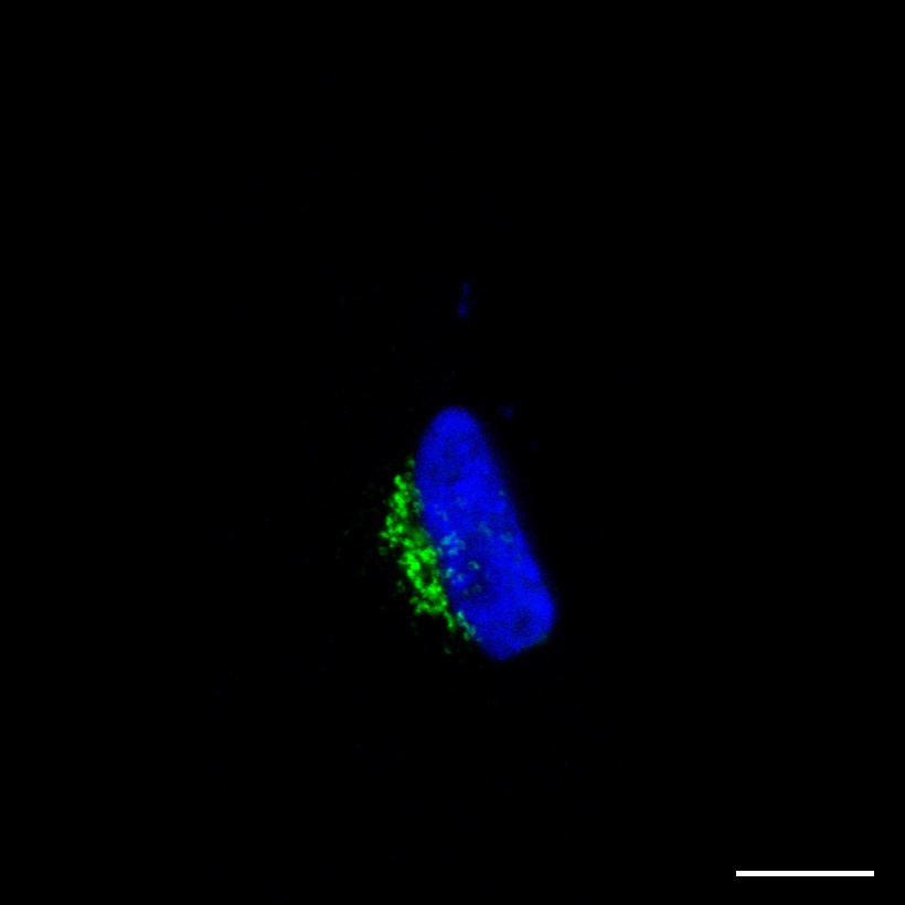
Staining of the Golgi apparatus in human cell line U-251 MG (HPA001677)
Scale bar represents 10µm
Golgi apparatus
The Golgi apparatus consists of several stacks of membrane cisternae with each of them containing a specific set of proteins. It is the key organelle in the secretory pathway: it receives newly synthesized proteins from the endoplasmic reticulum, modifies them and subsequently exports them to their final destination within or outside the cell.
Immunofluorescent staining
The Golgi apparatus is located next to the nucleus. Its morphology can vary between cell lines, but typically the cisternae are stacked in a ribbon-like form. Often it is spherically shaped and centered around the microtubule-organizing center, whereas in other cell lines it encircles the whole nucleus.