Renal cancer
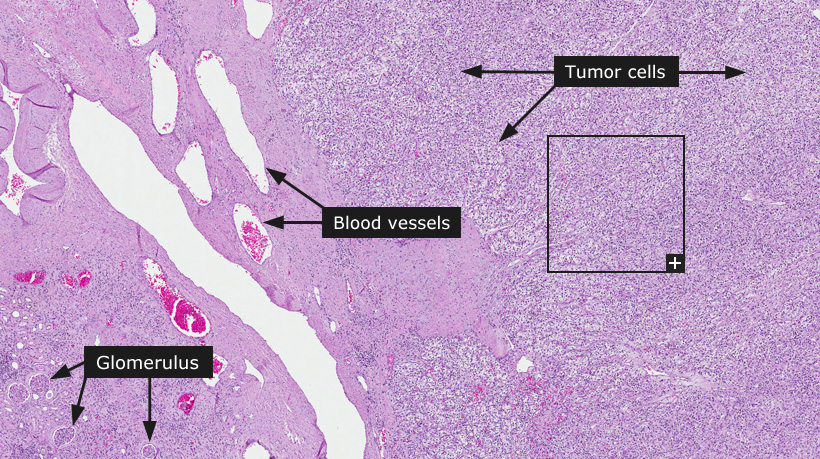
Female, 68 years, clear cell renal cell carcinoma 
Renal cancer
Renal cell carcinoma is almost exclusively a cancer of adults and it is two to three times more common in males than in females. The clinical course is highly unpredictable and recurrence ten years or more after the initial resection of a primary tumor is not uncommon. Several cases present as a metastatic carcinoma of unknown primary. Although smoking, industrial chemicals and obesity have been implicated as risk factors, in most cases the underlying carcinogenic influence is unknown. Clinical symptoms include hematuria, pain and mass in the flank as well as systemic symptoms such as fever, malaise and anemia.
Renal cell carcinoma consists of a family of carcinomas which are derived from the epithelium of renal tubules. The most frequent forms are clear cell renal cell carcinoma, papillary renal cell carcinoma, chromophobe renal cell carcinoma and collecting duct carcinoma. Approximately two thirds of all renal cell carcinomas are clear cell renal cell carcinomas and are signified by the appearance of tumor cells with abundant clear cytoplasm. Tumors can arise anywhere in the renal cortex and are typically surrounded by a fibrous pseudo-capsule.
Microscopically, clear cell renal cell carcinomas typically present a network of small blood vessels of uniform small caliber in addition to an abundant clear cytoplasm of tumor cells. The clarity of the cytoplasm is due to ample content of lipids and glycogen. Small and large cysts, hemorrhage and areas of necrosis are commonly found within the tumor. Of renal cell carcinomas, approximately 10-15% are papillary renal cell carcinomas, which are characterized by a predominantly papillary or tubulo-papillary growth pattern. Chromophobe renal cell carcinoma account for approximately 5% of all renal cell carcinomas and is the least aggressive variant. Chromophobe renal cell carcinomas are characterized by the presence of large numbers of minute intracytoplasmic vesicles with a flocculent appearance in tumor cells. All renal cell carcinomas can exhibit sarcomatoid changes. Benign tumors in the kidney also exist as adenomas, which are smaller tumors (less than 0.5 cm in diameter) compared to renal cell carcinomas, and oncocytomas which are characterized by tumor cells exhibiting large, intensely eosinophilic and finely granular cytoplasm and rounded nuclei.
The extent of spread of the primary tumor is the dominating factor that determines prognosis and is also the basis for tumor staging according to the TNM system. Renal cell carcinomas frequently invade the renal venous system and metastasize via the hematogenic route. Metastases in regional lymph nodes without hematogenic spread to distant organs are uncommon.
Normal tissue: Kidney
|