Fallopian tube

Fallopian tube
The normal fallopian tube extends from the area of its corresponding ovary to its terminus in the uterus. The tube measures between 9-11 cm in length. At the ovarian end, the tube opens into the peritoneal cavity and is composed of approximately 25 finger-like projections termed the fimbriae. In its extrauterine course, the fallopian tube is enveloped in a peritoneal fold along the superior margin of the broad ligament.
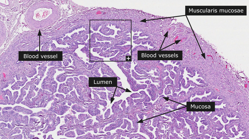
The fallopian tube is histologically composed of three layers: a mucosal membrane, a wall of smooth muscle and a serosal coat. The serosa is lined by flattened mesothelial cells. The muscularis mucosae is composed of two layers: an outer longitudinal and an inner circular layer. There is additionally, an inner longitudinal layer in the intramural segment of the tube that extends for 2 cm laterally. The inner circular layer forms the bulk of the muscular coat. The outer longitudinal layer comprises inconspicuous smooth muscle cells interspersed with loose connective tissue.
The mucosa rests directly on the muscularis. It consists of a luminal epithelial lining and a scant underlying lamina propria with sparse spindle and angulated cells. The luminal complexity is more marked towards the ovarian end compared to the interstitial and isthmic portions containing only five to six blunt papillae.
Three histologic cell types comprise the epithelial layer: ciliated (20-30%), secretory (55-60%) and intercalary cells. Ciliated cells are believed to be more frequent in the ovarian end of the fallopian tube. The ciliated cell has a columnar shape and contains a oval or round nucleus, often located perpendicular or parallel to the long axis of the cell. The secretory cell is usually a more narrow columnar cell with approximately the same height as the ciliated cell. The nucleus is ovoid and perpendicular to the long axis of the cell. The chromatin is more dense and the nucleolus smaller than that seen in the ciliated cell. The intercalary, or peg cell is a columnar cell occupied chiefly by a thin, dark-staining nucleus.
|