TISSUE
CELL
CANCER
ANTIBODY INFORMATION
Antibody HPA035809
Antibody HPA035810
Antibody HPA061496
Provider
Product name
Host species
Clonality
Purity
Other gene match
Released in version
References
VALIDATION SUMMARY
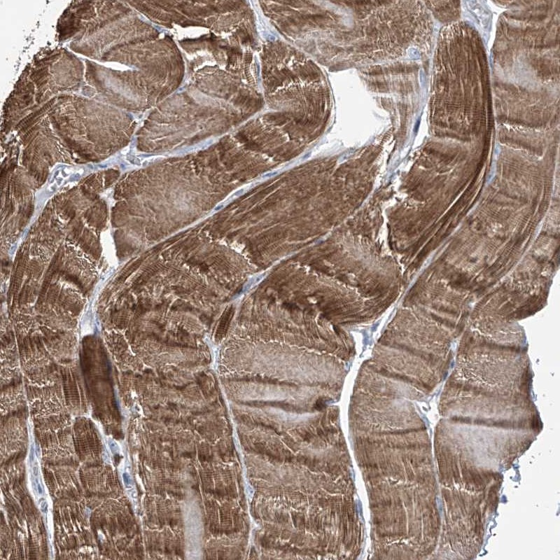
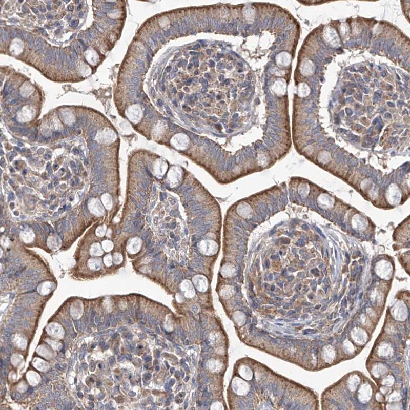
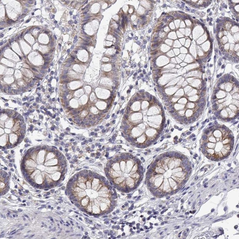
IMMUNOHISTOCHEMISTRY
Formal validation: Independent
Figure description
Standard validation
Expression
Retrieval
Antibody dilution
Literature conformity
RNA consistency
WESTERN BLOT
Target mass (kDa)
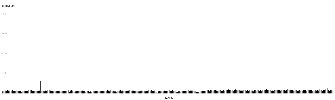
PROTEIN ARRAY
ANTIGEN INFORMATION
Antigen
Length (aa)
Antigen sequence
HLLREVEGKSVQSNLELLTQAKSMHHKYQNLKCPELFSFRLTKYGFSLPP
QYSGLDRIIKPFQVDVILDLNTAHPQ
SYHREILEGSLEPLRNNIERVEKVIILQGSKSVELKKKVEYKREEINSEF
EQIRLFLQNEQEMILRQIQDEEMNILAKLNENLVELSDYVSTL
VGNKPKWILGVCQDCLLRNWQDQPSVLGGFWAIGRYMKSGYVASGPKTTQ
LLPVVKPSK
Matching transcripts