TISSUE
CELL
CANCER
ANTIBODY INFORMATION
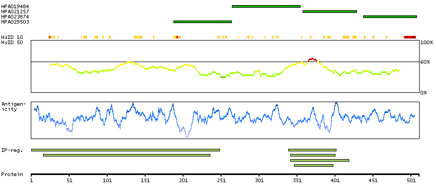
Antibody HPA019484
Antibody HPA021257
Antibody HPA023874
Antibody HPA029503
Provider
Product name
Host species
Clonality
Purity
Other gene match
Released in version
References
VALIDATION SUMMARY
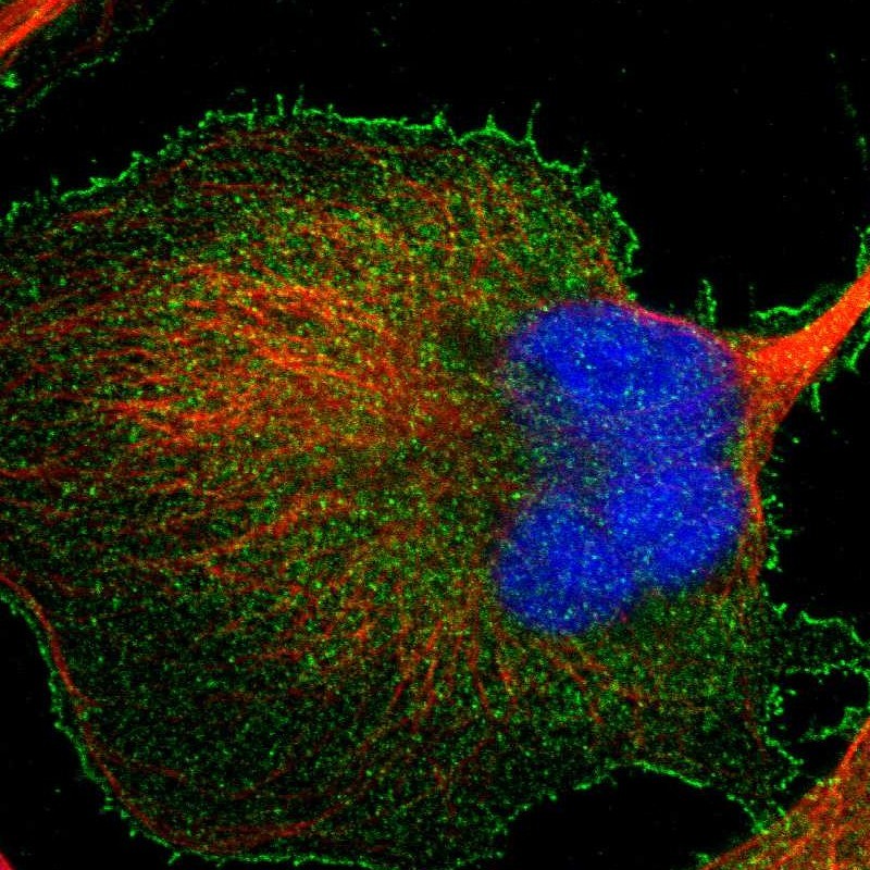
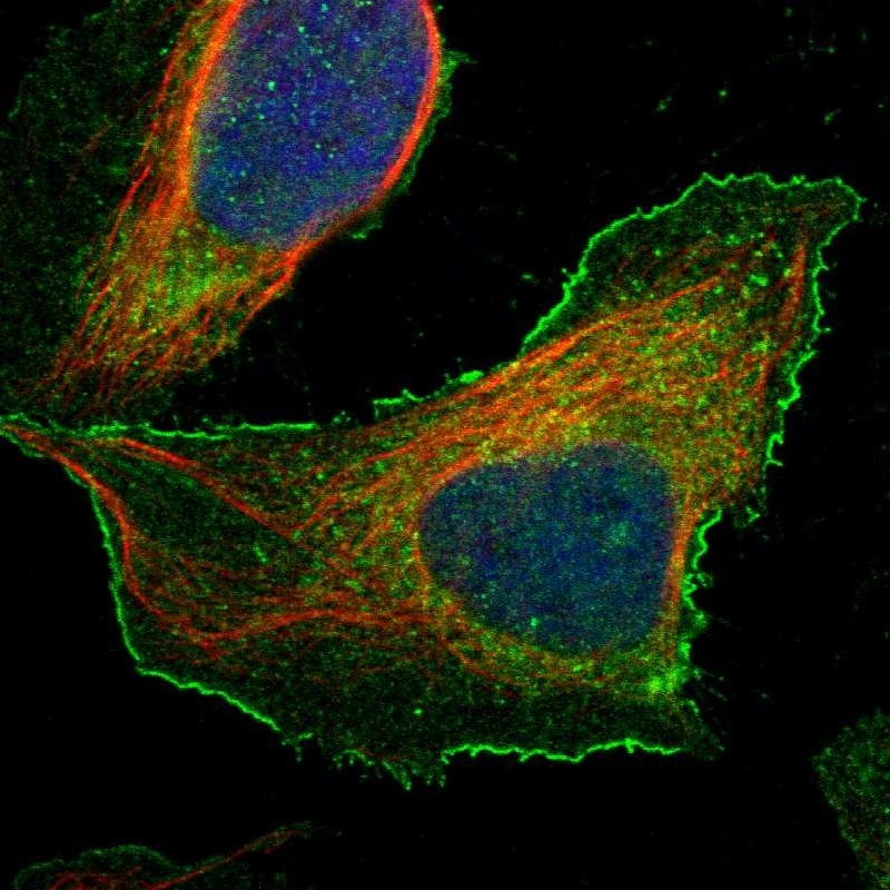
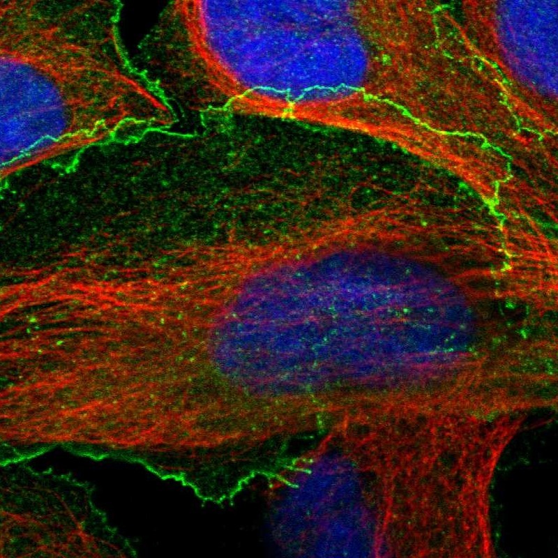
IMMUNOCYTOCHEMISTRY
Formal validation: Independent
Standard validation
Figure description
Antibody dilution
Literature conformity
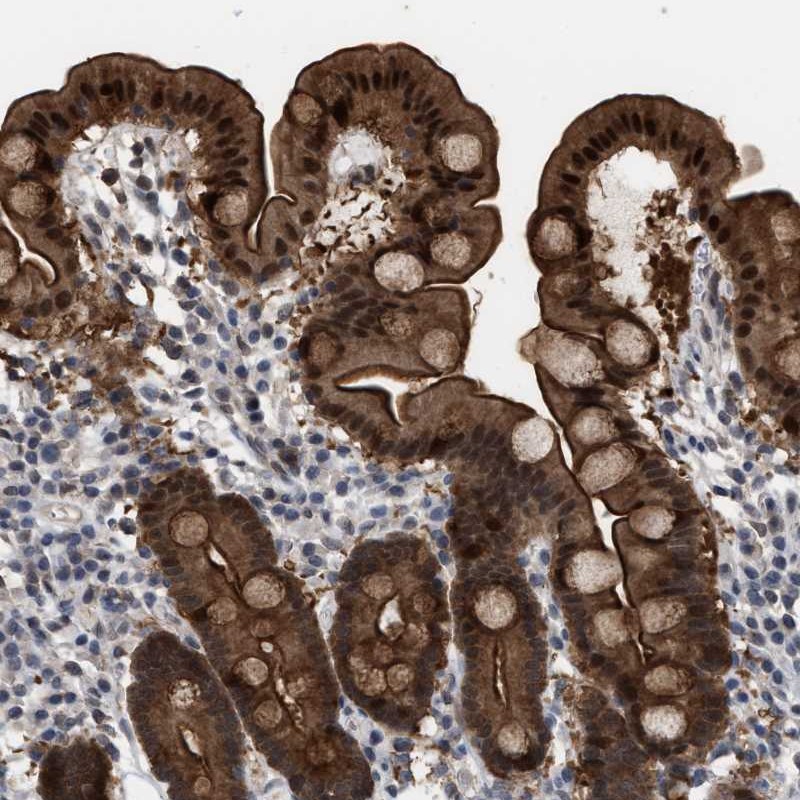
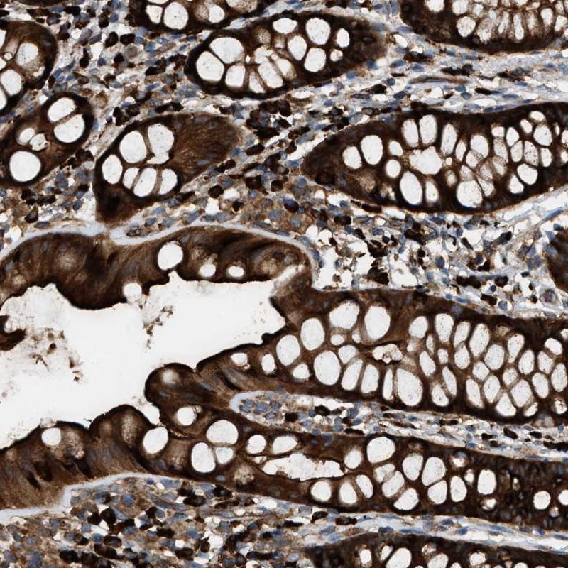
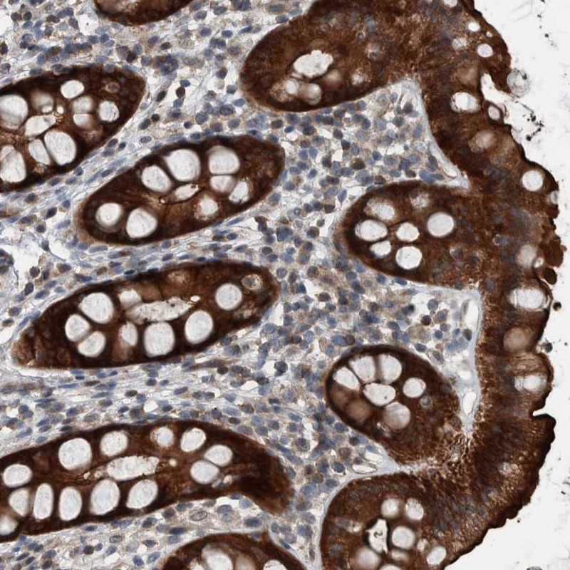
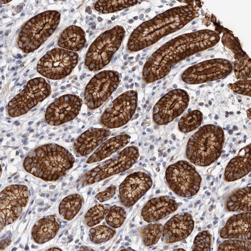
IMMUNOHISTOCHEMISTRY
Formal validation: Orthogonal
Expression
Retrieval
RNA consistency
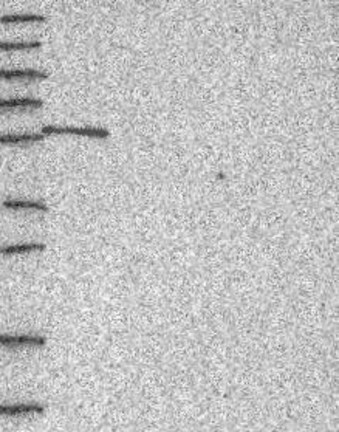
WESTERN BLOT
Target mass (kDa)
PROTEIN ARRAY
ANTIGEN INFORMATION
Antigen
Length (aa)
Antigen sequence
ERSNVVRKDYDTLSKCSPKMPPAPSGRAYTSPLIDMFNNPATAAPNSQRV
NNSTGTSEDPSLQRSVSVATGLNMMKKQKVKTIFPHTAGSN
LLSFAQGDVITLLIPEEKDGWLYGEHDVSKARGWFPSSYTKLLEENETEA
VTVPTPSPTPVRSISTVNLSEN
DYLECLSMGAAADRRADSARTTSTFKAPASKPETAAPNDANGTAKPPFLS
GENPFATVKLRPTVTNDRSAP
EEKRRFCFLVDKHCGFANHIHYYHLQSAELLNSKLPRWQETCVDAIKVPE
KIMNMIEEIKTPASTPVSGTPQASPMI
Matching transcripts