TISSUE
CELL
CANCER
ANTIBODY INFORMATION
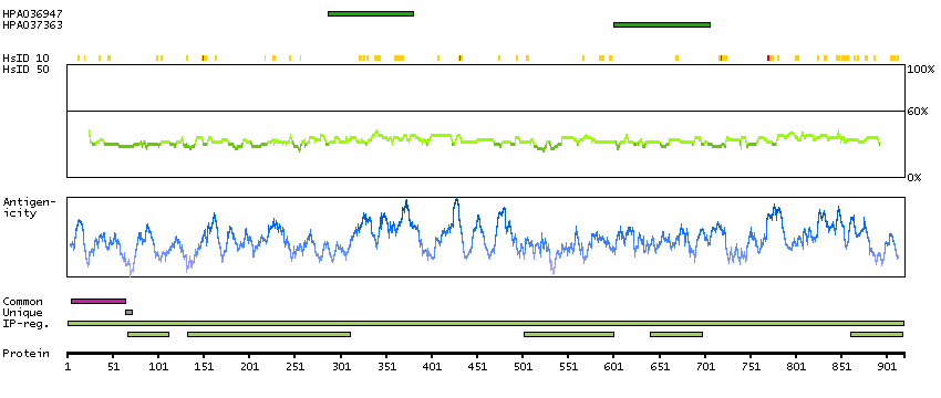
Antibody HPA036947
Antibody HPA037363
Antibody CAB012423
Provider
Product name
Host species
Clonality
Purity
Other gene match
Released in version
References
VALIDATION SUMMARY
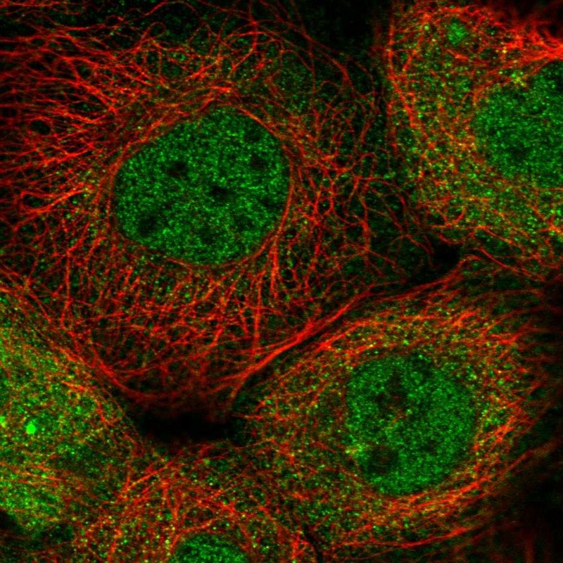
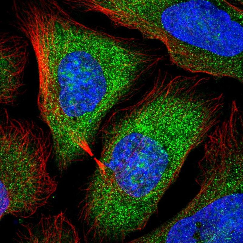
IMMUNOCYTOCHEMISTRY
Standard validation
Figure description
Antibody dilution
Literature conformity
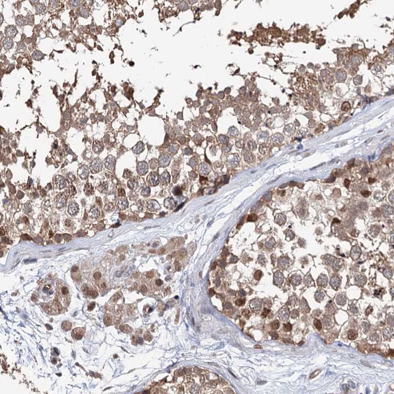
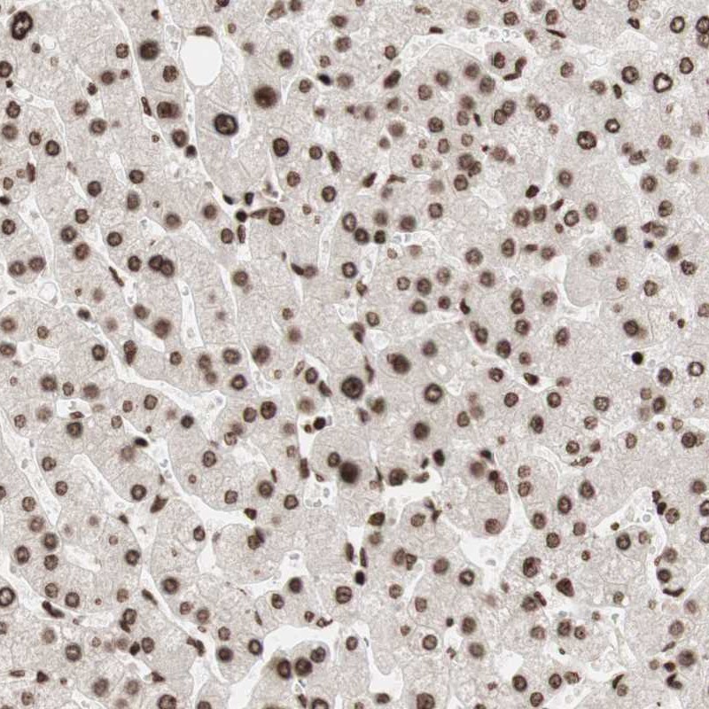
IMMUNOHISTOCHEMISTRY
Formal validation: Independent
Expression
Retrieval
RNA consistency
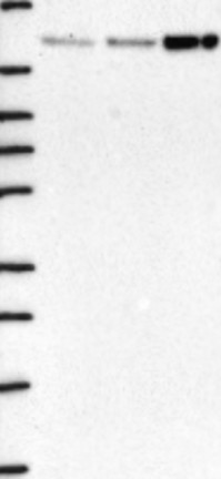
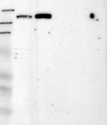
WESTERN BLOT
Formal validation: Genetic
Target mass (kDa)
Loading control
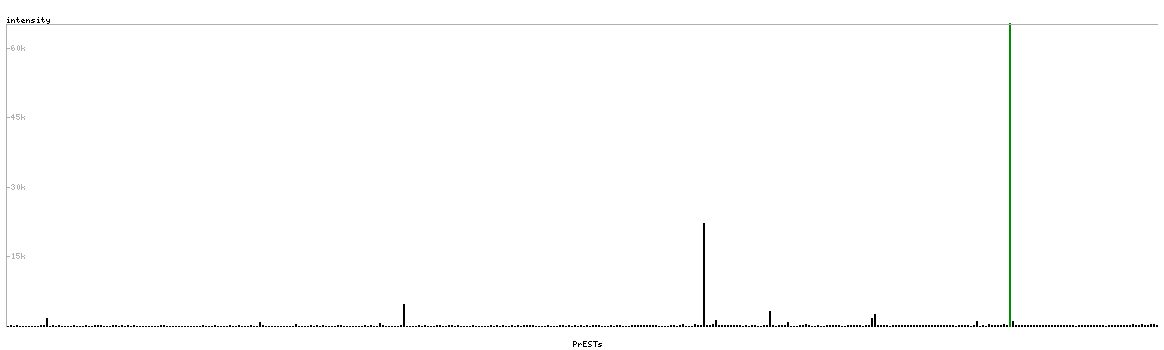
PROTEIN ARRAY
ANTIGEN INFORMATION
Antigen
Length (aa)
Antigen sequence
DKSVLVCKNAIQLLASFLANNPFSCKLSDADLAGPLQKETQKLQEMRAQR
RTAAASAVLDPEEEWEAMLPELKSTLQQLLQLPQGEEEIPEQIA
VGTIQCLEEILCEFVQKDELKPAVTQLLWERATEKVACCPLERCSSVMLL
GMMARGKPEIVGSNLDTLVSIGLDEKFPQDYRLAQQVCHAIANISDRRKP
SLGKRH
Matching transcripts