TISSUE
CELL
CANCER
ANTIBODY INFORMATION
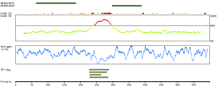
Antibody HPA023871
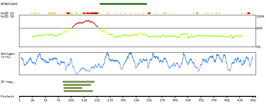
Antibody HPA052645
Provider
Product name
Host species
Clonality
Purity
Other gene match
Released in version
References
VALIDATION SUMMARY
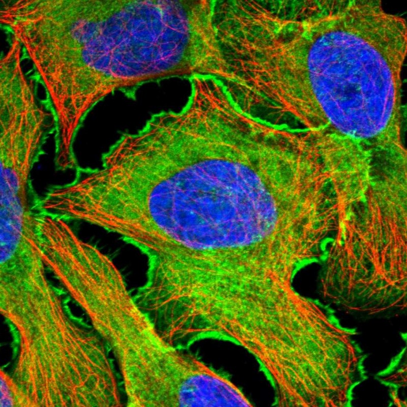
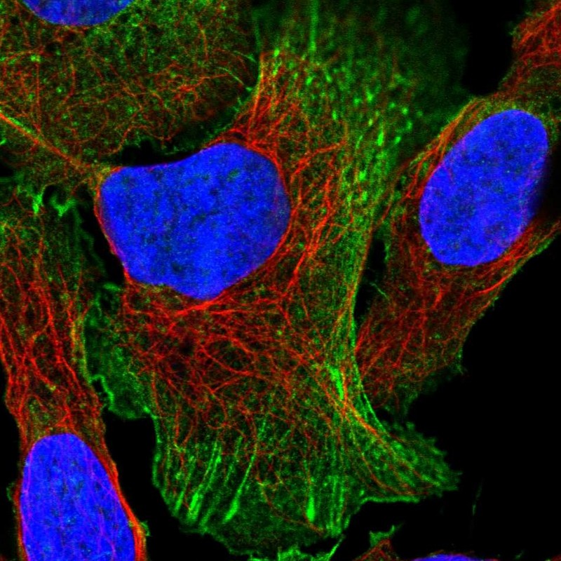
IMMUNOCYTOCHEMISTRY
Formal validation: Independent
Standard validation
Figure description
Antibody dilution
Literature conformity
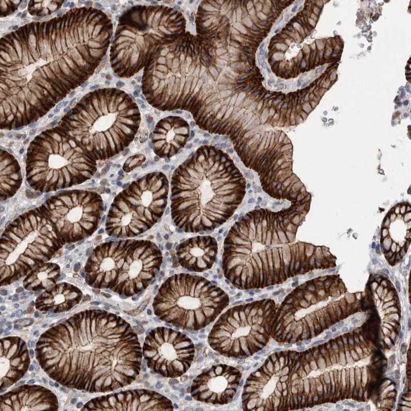
IMMUNOHISTOCHEMISTRY
Formal validation: Orthogonal
Expression
Retrieval
RNA consistency
WESTERN BLOT
Target mass (kDa)
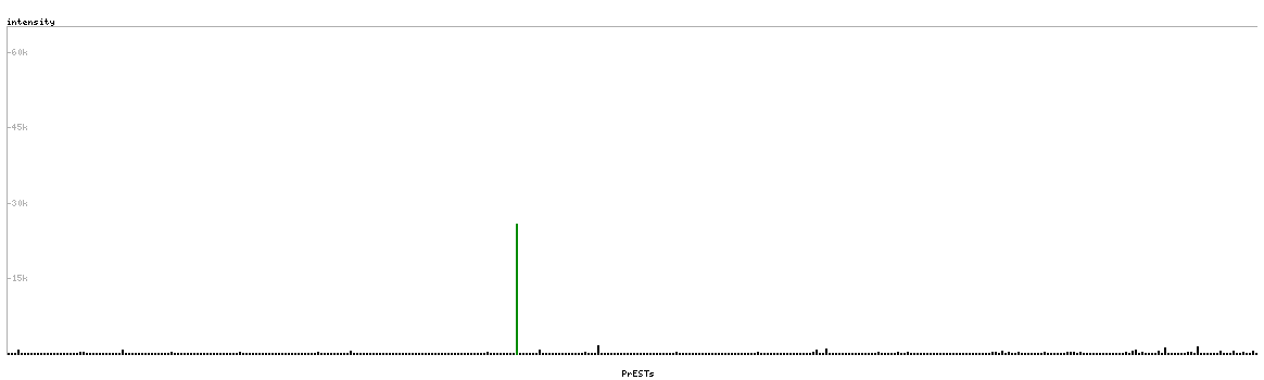
PROTEIN ARRAY
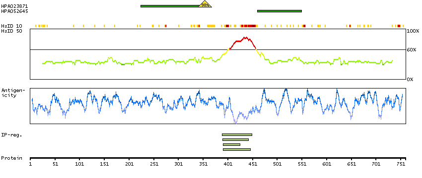
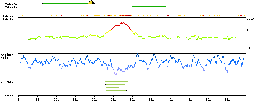
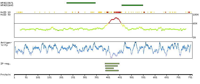
ANTIGEN INFORMATION
Antigen
Length (aa)
Antigen sequence
ISENSYSLDDLEIGPGQLSSSTFDSEKNESRRNLELPRLSETSIKDRMAK
YQAAVSKQSSSTNYTNELKASGGEIKIHKMEQKENVPPGPEVCITHQEGE
KISANENSLAVRSTPAEDDSRDS
RPHKDLWASKNENEEILERPAQLANARETPHSPGVEDAPIAKVGVLAASM
EAKASSQQEKEDKPAETKKLRIAWPPPTELGSSGSALEEGI
Matching transcripts
RELEVANT PUBLICATIONS
1.