TISSUE
CELL
CANCER
ANTIBODY INFORMATION
Antibody HPA041943
Antibody HPA043562
Antibody CAB010910
Provider
Product name
Host species
Clonality
Purity
Other gene match
Released in version
References
VALIDATION SUMMARY
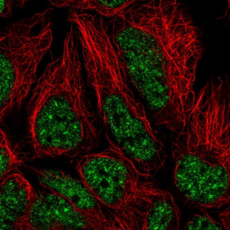
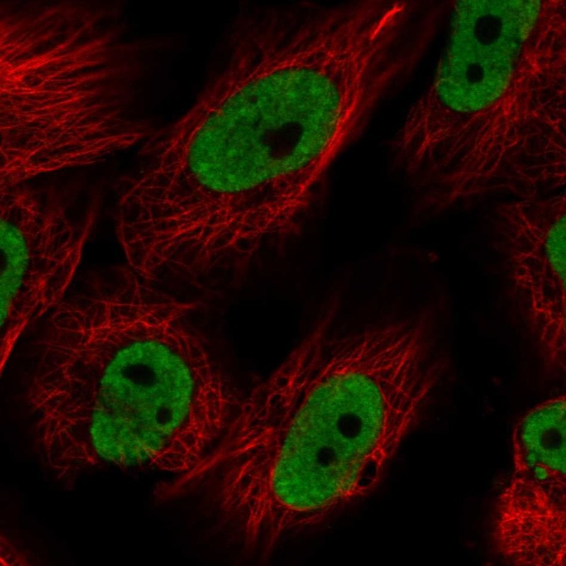
IMMUNOCYTOCHEMISTRY
Standard validation
Figure description
Antibody dilution
Literature conformity
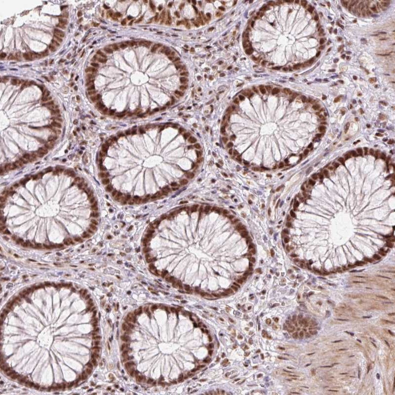
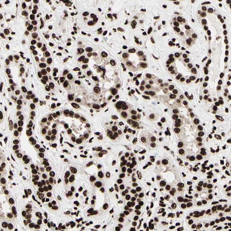
IMMUNOHISTOCHEMISTRY
Expression
Retrieval
RNA consistency
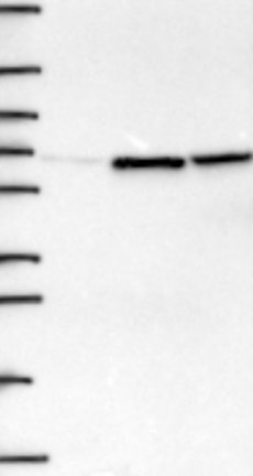
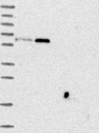
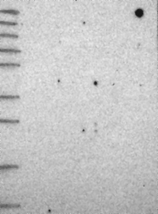
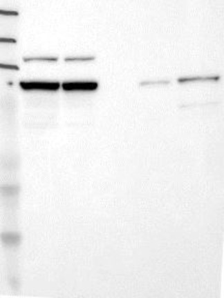
WESTERN BLOT
Formal validation: Genetic
Target mass (kDa)
Loading control
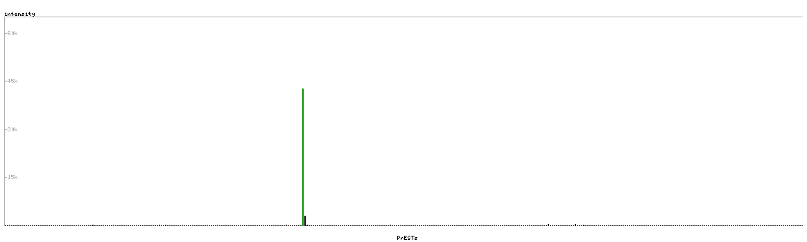
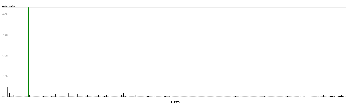
PROTEIN ARRAY
ANTIGEN INFORMATION
Antigen
Length (aa)
Antigen sequence
LTRGAKEEHGGLIRSPRHEKKKKVRKYWDVPPPGFEHITPMQYKAMQAAG
QIPATALLPTMTPDGLAVTPTPVPVVGSQMTRQARRLYVGNIPFGITEEA
PDSAHKLFIGGLPNYLNDDQVKELLTSFGPLKAFNLVKDSATGLSKGYAF
CEYVDINVTDQAIAGLNGMQLGDKKLLVQRASVGAKNATLVS
Matching transcripts