TISSUE
CELL
CANCER
ANTIBODY INFORMATION
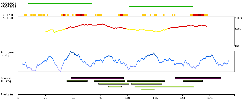
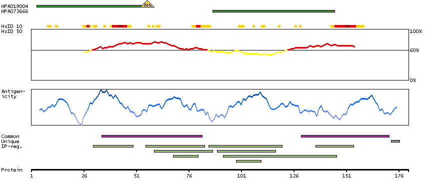
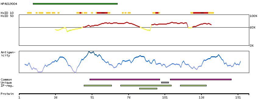
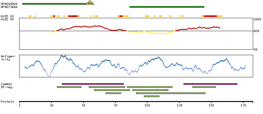
Antibody HPA019004
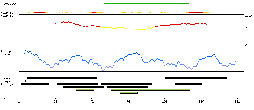
Antibody HPA073666
Provider
Product name
Host species
Clonality
Purity
Other gene match
Released in version
References
VALIDATION SUMMARY
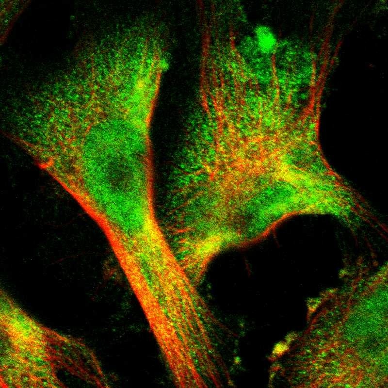
IMMUNOCYTOCHEMISTRY
Standard validation
Figure description
Antibody dilution
Literature conformity
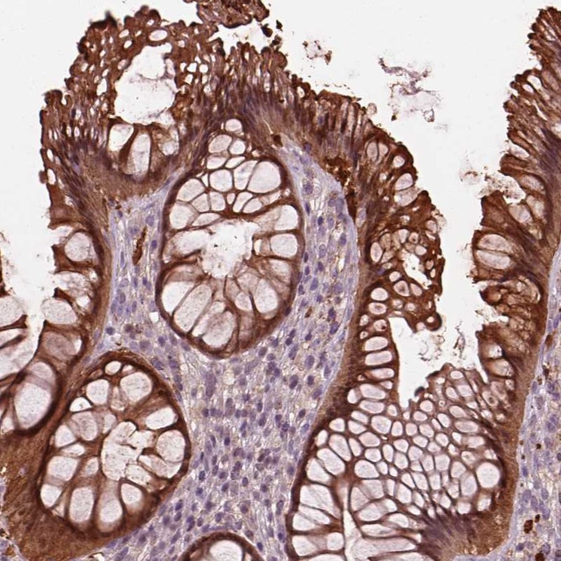
IMMUNOHISTOCHEMISTRY
Formal validation: Orthogonal
Formal validation: Independent
Expression
Retrieval
RNA consistency
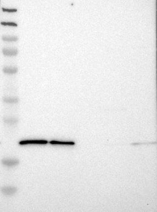
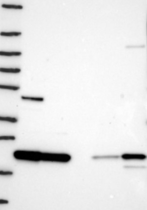
WESTERN BLOT
Target mass (kDa)
PROTEIN ARRAY
ANTIGEN INFORMATION
Antigen
Length (aa)
Antigen sequence
GGGYYPGGYGGAPGGPAFPGQTQDPLYGYFAAVAGQDGQIDADELQRCLT
QSGIAGGYK
LNGWRQHFISFDTDRSGTVDPQELQKALTTMGFRLSPQAVNSIAKRYSTN
GKITFDDYI
Matching transcripts