We use cookies to enhance the usability of our website. If you continue, we'll assume that you are happy to receive all cookies. More information. Don't show this again.
Spearman correlation >0.6 for protein expression in tissues using independent antibodies.
Validated
Spearman correlation >0.6 for protein expression in tissues using independent antibodies.
Figure description
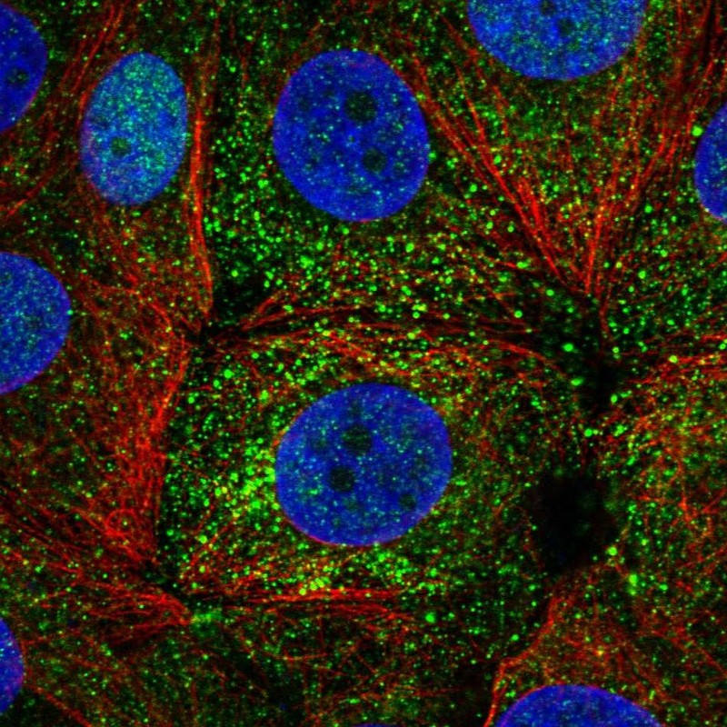
Distribution of protein expression (antibody staining). Spearman correlation with HPA000450 across 74 cell types.
Distribution of protein expression (antibody staining). Spearman correlation with HPA000449 across 74 cell types.
Standard validation
Supported
Supported
Supported
Supported
Supported
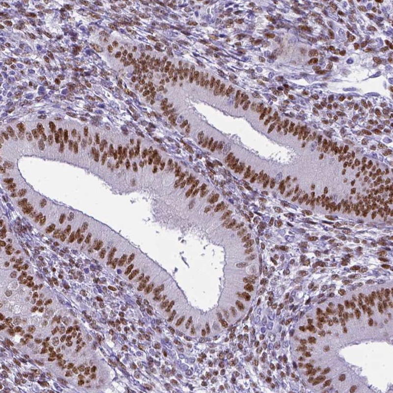
Figure description
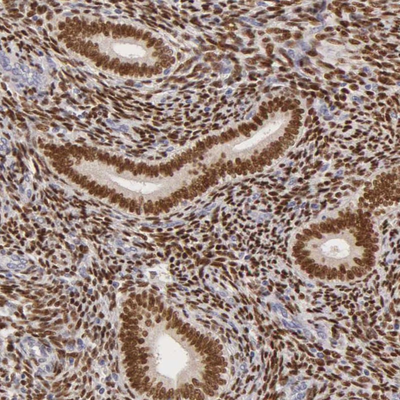
Immunohistochemical staining of human uterus shows strong nuclear positivity in both stroma and glandular cells.
Immunohistochemical staining of human corpus, uterine shows strong nuclear positivity in glandular cells and stromal cells.
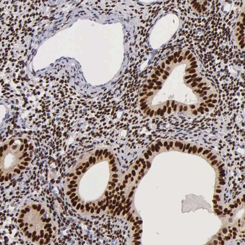
Immunohistochemical staining of human corpus, uterine shows strong nuclear positivity in glandular cells.
Immunohistochemical staining of human uterus, pre-menopause shows strong cytoplasmic positivity in glandular cells.
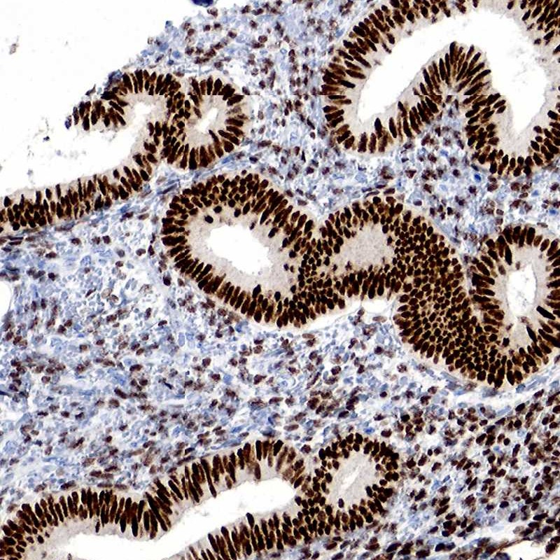
Immunohistochemical staining of human endometrium shows nuclear positivity with moderate intensity in glandular cells and with weak intensity in cells of endometrial stroma.
Expression
RNA: detected in 23 tissues Protein: detected in 12 cell types
RNA: detected in 23 tissues Protein: detected in 25 cell types
RNA: detected in 23 tissues Protein: detected in 10 cell types
RNA: detected in 23 tissues Protein: detected in 13 cell types
RNA: detected in 23 tissues Protein: detected in 11 cell types
Retrieval
HIER pH6
HIER pH6
HIER pH9
HIER pH6
HIER pH6
Antibody dilution
1:200
1:100
1:150
1:150
1:1000
Literature conformity
Consistent with extensive gene/protein characterization data.
Consistent with extensive gene/protein characterization data.
Consistent with extensive gene/protein characterization data.
Consistent with extensive gene/protein characterization data.
Consistent with extensive gene/protein characterization data.
RNA consistency
Consistent with RNA expression data.
Mainly consistent with RNA expression data.
Mainly consistent with RNA expression data.
Consistent with RNA expression data.
Consistent with RNA expression data.
WESTERN BLOT
Antibody HPA000449
Antibody HPA000450
Antibody CAB000037
Antibody CAB055099
Antibody CAB072858
Standard validation
Supported
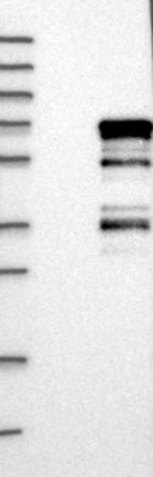
Band of predicted size in kDa (+/-20%) with additional bands present.
Supported
Band of predicted size in kDa (+/-20%) with additional bands present.
Uncertain
Analysis performed using a standard panel of samples. No bands detected.
Uncertain
Analysis performed using a standard panel of samples. Weak band of predicted size but with additional bands of higher intensity also present.
Uncertain
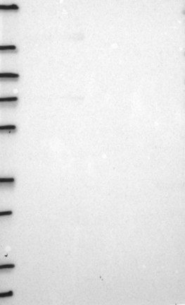
Analysis performed using a standard panel of samples. No bands detected.
Figure description
Lane 1: Marker [kDa] 250, 130, 95, 72, 55, 36, 28, 17, 10 Lane 2: Negative control (vector only transfected HEK293T lysate) Lane 3: Over-expression Lysate (Co-expressed with a C-terminal myc-DDK tag (~3.1 kDa) in mammalian HEK293T cells, LY400046)
Lane 1: Marker [kDa] 250, 130, 95, 72, 55, 36, 28, 17, 10 Lane 2: Negative control (vector only transfected HEK293T lysate) Lane 3: Over-expression Lysate (Co-expressed with a C-terminal myc-DDK tag (~3.1 kDa) in mammalian HEK293T cells, LY400046)
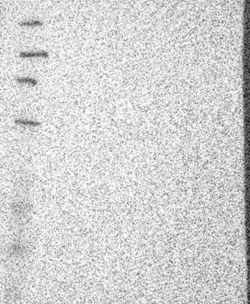
Lane 1: Marker [kDa] 219, 112, 85, 49, 32, 25, 17.5 Lane 2: RT4 Lane 3: U-251 MG Lane 4: A-431 Lane 5: Liver Lane 6: Tonsil
Lane 1: Marker [kDa] 250, 130, 95, 72, 55, 36, 28, 17, 10 Lane 2: RT4 Lane 3: U-251 MG Lane 4: Human Plasma Lane 5: Liver Lane 6: Tonsil
Target mass (kDa)
66.2
66.2, 36.7
66.2, 36.7, 35.3, 16.3, 12.3, 12.1, 9
66.2, 36.7, 35.3, 16.3, 12.3, 12.1, 9
66.2, 36.7, 35.3, 16.3, 12.3, 12.1, 9
Antibody dilution
1:250
1:250
1:500
1:500
1:1000
PROTEIN ARRAY
Antibody HPA000449
Antibody HPA000450
Antibody CAB000037
Antibody CAB055099
Antibody CAB072858
Standard validation
Supported
Pass with single peak corresponding to interaction only with its own antigen.
Supported
Pass with single peak corresponding to interaction only with its own antigen.
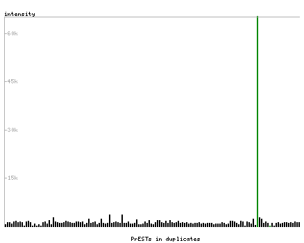
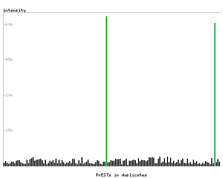
Figure description
Antibody specificity analysis with protein arrays. Predicted and matching interactions are shown in green.
Antibody specificity analysis with protein arrays. Predicted and matching interactions are shown in green.