We use cookies to enhance the usability of our website. If you continue, we'll assume that you are happy to receive all cookies. More information. Don't show this again.
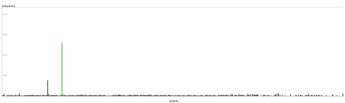
Cytoplasmic region of segmented cells in 10x-images
Antibody dilution
1:96
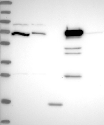
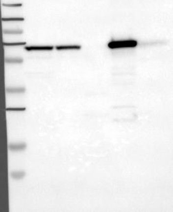
Formal validation: Tagged protein
Validated
Antibody staining overlaps with GFP tagged protein
Validated
Antibody staining overlaps with GFP tagged protein
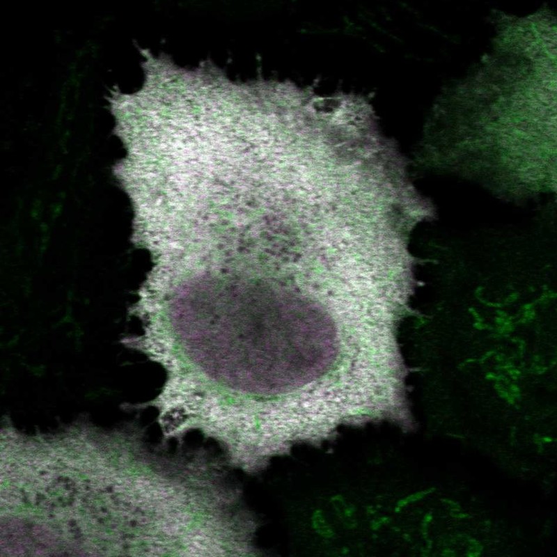
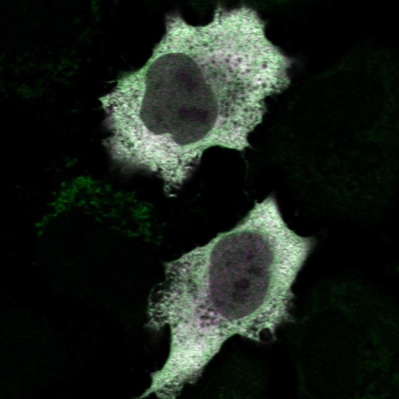
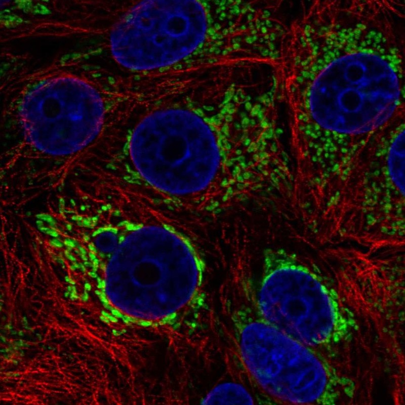
Figure description
Immunofluorescent staining of transgenic HeLa cells show antibody staining in cytoplasm, mitochondria & nucleus but excluded from the nucleoli and GFP expression in cytoplasm & nucleus but excluded from the nucleoli.
Immunofluorescent staining of transgenic HeLa cells show antibody staining in cytoplasm & mitochondria and GFP expression in cytoplasm & nucleus but excluded from the nucleoli.
Antibody dilution
1:96
1:200
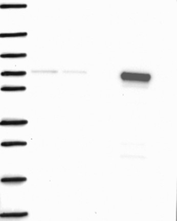
Formal validation: Independent
Validated
Antibody staining overlaps with antibody HPA053502.
Validated
Antibody staining overlaps with antibody HPA051162.
Standard validation
Supported
Supported
Supported
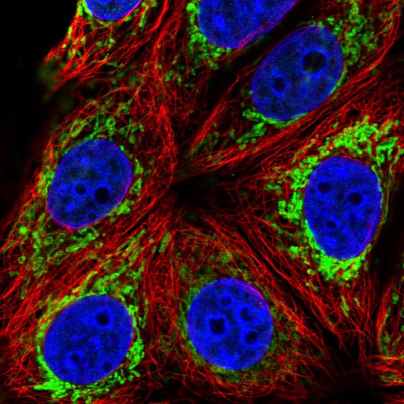
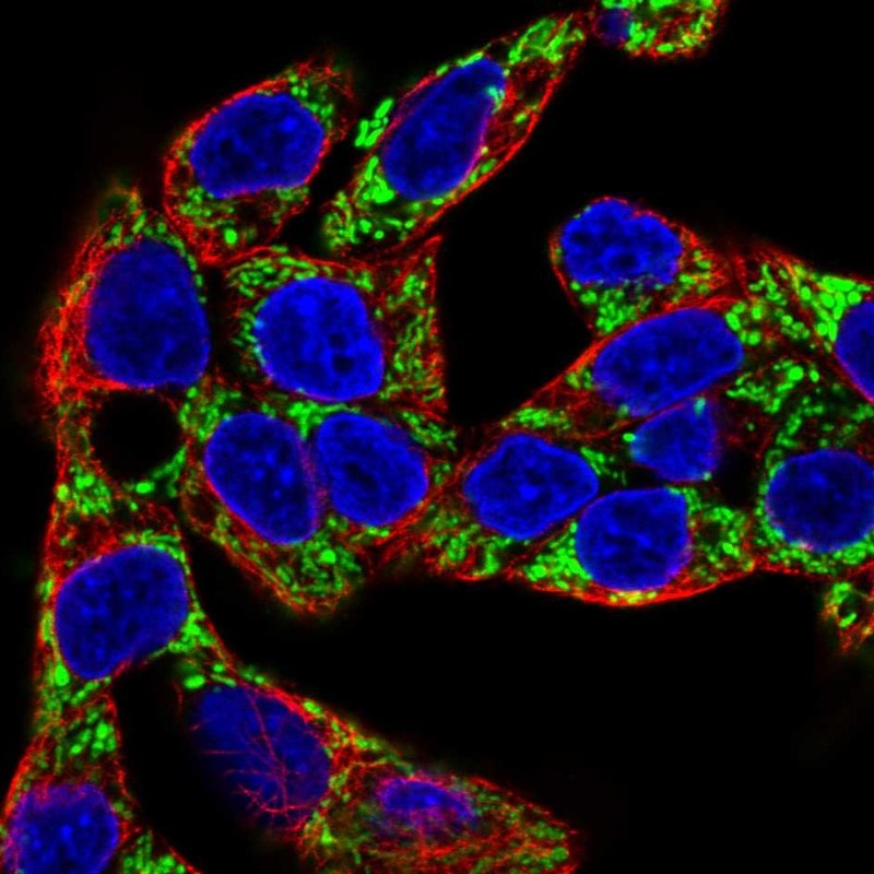
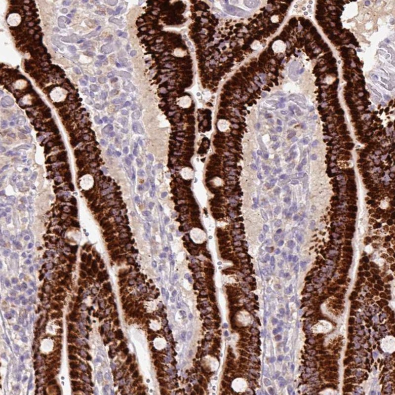
Figure description
Immunofluorescent staining of human cell line MCF7 shows localization to mitochondria.
Immunofluorescent staining of human cell line Hep G2 shows localization to mitochondria.
Immunofluorescent staining of human cell line MCF7 shows localization to mitochondria.
Antibody dilution
1:96
1:200
1:188
Literature conformity
The subcellular location is supported by literature.
The subcellular location is supported by literature.
The subcellular location is supported by literature.
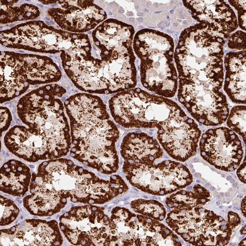
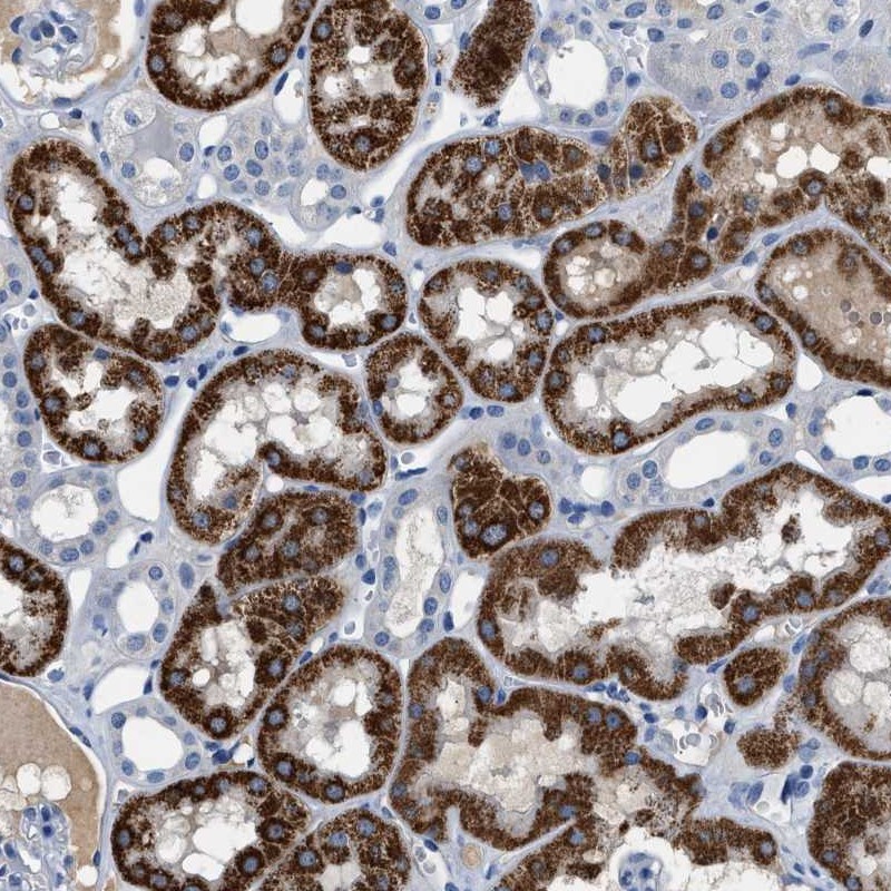
IMMUNOHISTOCHEMISTRY
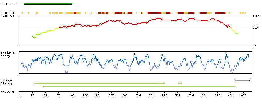
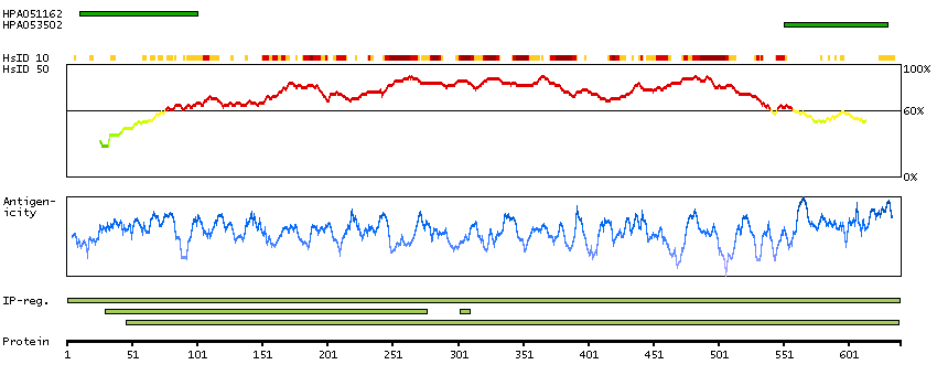
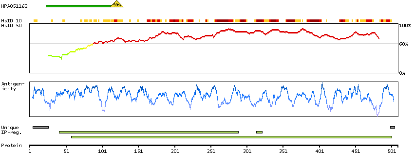
Antibody HPA051162
Antibody HPA053502
Antibody CAB018734
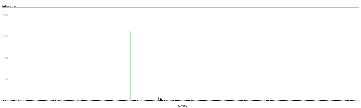
Formal validation: Orthogonal
Validated
Pearson correlation >0.6 for protein and RNA expression in cell lines.