We use cookies to enhance the usability of our website. If you continue, we'll assume that you are happy to receive all cookies. More information. Don't show this again.
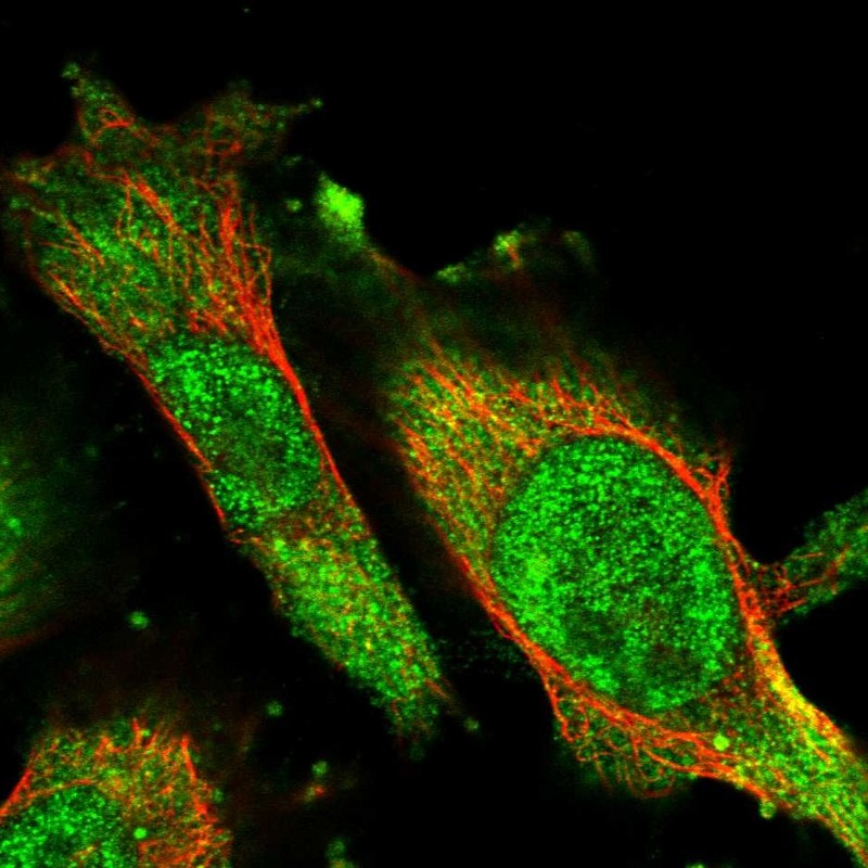
Immunofluorescent staining of human cell line U-251 MG shows localization to nucleoplasm & cytosol.
Antibody dilution
1:10
Literature conformity
The subcellular location is supported by literature.
IMMUNOHISTOCHEMISTRY
Antibody HPA025736
Antibody HPA025765
Antibody HPA025811
Antibody CAB021106
Formal validation: Independent
Validated
Pearson correlation >0.6 for protein expression in cell lines using independent antibodies.
Validated
Pearson correlation >0.6 for protein expression in cell lines using independent antibodies.
Validated
Pearson correlation >0.6 for protein expression in cell lines using independent antibodies.
Figure description
Distribution of protein expression (antibody staining). Pearson correlation with HPA025811 across 45 cell lines. Pearson correlation with HPA025765 across 46 cell lines.
Distribution of protein expression (antibody staining). Pearson correlation with HPA025811 across 45 cell lines. Pearson correlation with HPA025736 across 46 cell lines.
Distribution of protein expression (antibody staining). Pearson correlation with HPA025765 across 45 cell lines. Pearson correlation with HPA025736 across 45 cell lines.
Formal validation: Independent
Validated
Spearman correlation >0.6 for protein expression in tissues using independent antibodies.
Validated
Spearman correlation >0.6 for protein expression in tissues using independent antibodies.
Validated
Spearman correlation >0.6 for protein expression in tissues using independent antibodies.
Figure description
Distribution of protein expression (antibody staining). Spearman correlation with HPA025811 across 75 cell types. Spearman correlation with HPA025765 across 75 cell types.
Distribution of protein expression (antibody staining). Spearman correlation with HPA025811 across 76 cell types. Spearman correlation with HPA025736 across 75 cell types.
Distribution of protein expression (antibody staining). Spearman correlation with HPA025765 across 76 cell types. Spearman correlation with HPA025736 across 75 cell types.
Standard validation
Supported
Supported
Supported
Supported
Figure description
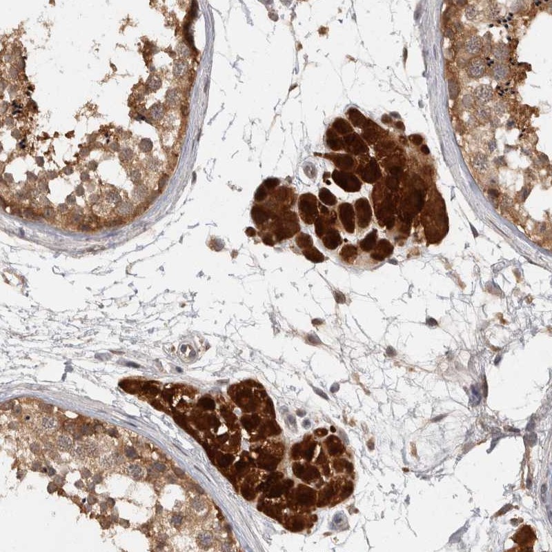
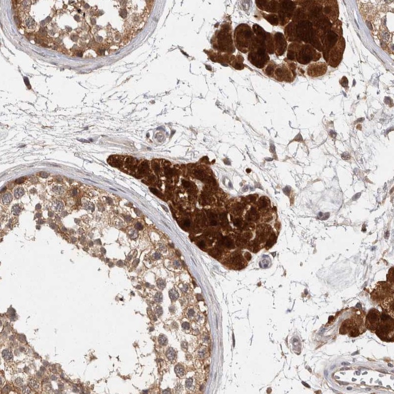
Immunohistochemical staining of human testis shows strong cytoplasmic positivity in leydig cells.
Immunohistochemical staining of human testis shows strong cytoplasmic positivity in leydig cells.
Immunohistochemical staining of human testis shows strong cytoplasmic positivity in leydig cells.
Immunohistochemical staining of human small intestine shows cytoplasmic positivity in glandular cells.
Expression
RNA: detected in 35 tissues Protein: detected in 72 cell types
RNA: detected in 35 tissues Protein: detected in 68 cell types
RNA: detected in 35 tissues Protein: detected in 65 cell types
RNA: detected in 35 tissues Protein: detected in 66 cell types
Retrieval
HIER pH6
HIER pH6
HIER pH6
HIER pH6
Antibody dilution
1:45
1:65
1:10
1:12000
Literature conformity
Consistent with extensive gene/protein characterization data.
Consistent with extensive gene/protein characterization data.
Consistent with extensive gene/protein characterization data.
Consistent with extensive gene/protein characterization data.
RNA consistency
Mainly consistent with RNA expression data.
Mainly consistent with RNA expression data.
Mainly consistent with RNA expression data.
Mainly consistent with RNA expression data.
WESTERN BLOT
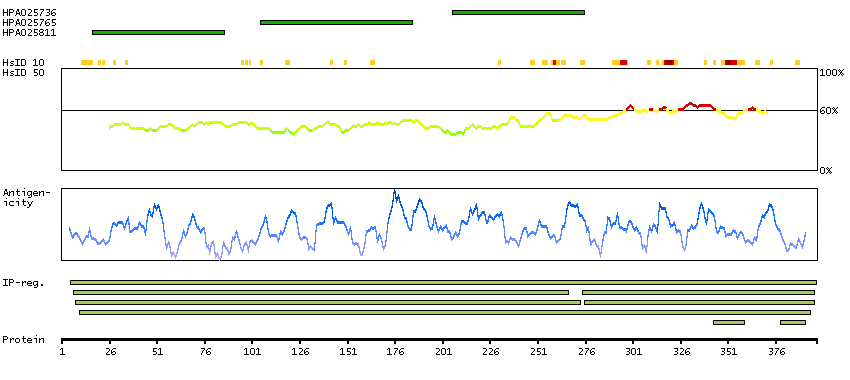
Antibody HPA025736
Antibody HPA025765
Antibody HPA025811
Antibody CAB021106
Standard validation
Uncertain
Analysis performed using a standard panel of samples. Weak band of predicted size but with additional bands of higher intensity also present.
Supported
Analysis performed using a standard panel of samples. Single band corresponding to the predicted size in kDa (+/-20%).
Supported
Analysis performed using a standard panel of samples. Single band corresponding to the predicted size in kDa (+/-20%).
Uncertain
Analysis performed using a standard panel of samples. Only bands not corresponding to the predicted size.
Figure description
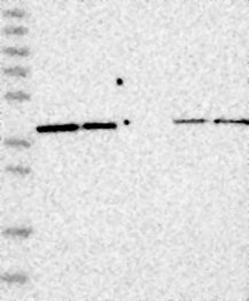
Lane 1: Marker [kDa] 230, 130, 95, 72, 56, 36, 28, 17, 11 Lane 2: RT4 Lane 3: U-251 MG Lane 4: Human Plasma Lane 5: Liver Lane 6: Tonsil
Lane 1: Marker [kDa] 230, 130, 95, 72, 56, 36, 28, 17, 11 Lane 2: RT4 Lane 3: U-251 MG Lane 4: Human Plasma Lane 5: Liver Lane 6: Tonsil
Target mass (kDa)
41.4
41.4
41.4
41.4
Antibody dilution
1:250
1:250
1:250
1:1000
PROTEIN ARRAY
Antibody HPA025736
Antibody HPA025765
Antibody HPA025811
Antibody CAB021106
Standard validation
Supported
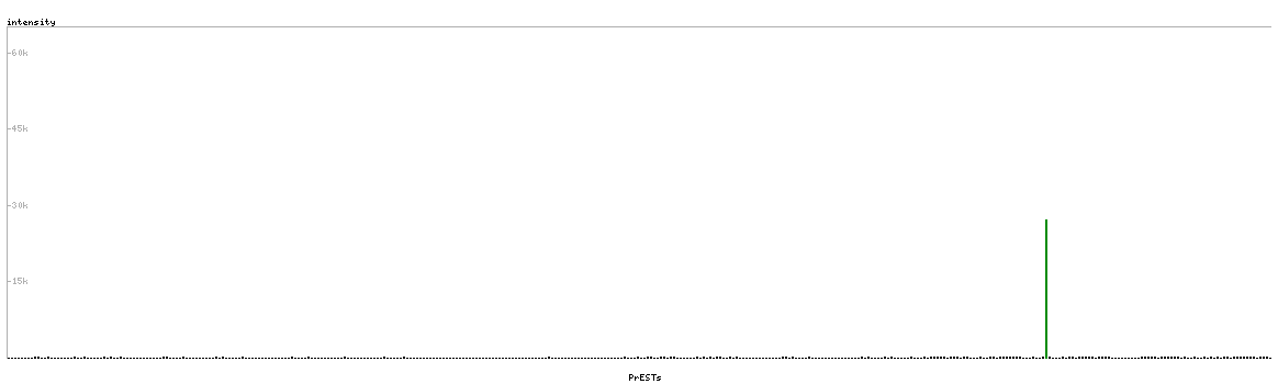
Pass with single peak corresponding to interaction only with its own antigen.
Supported
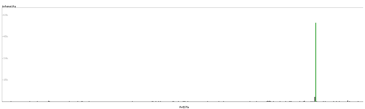
Pass with single peak corresponding to interaction only with its own antigen.
Supported
Pass with single peak corresponding to interaction only with its own antigen.
Figure description
Antibody specificity analysis with protein arrays. Predicted and matching interactions are shown in green.
Antibody specificity analysis with protein arrays. Predicted and matching interactions are shown in green.
Antibody specificity analysis with protein arrays. Predicted and matching interactions are shown in green.