TISSUE
CELL
CANCER
ANTIBODY INFORMATION
Antibody HPA020559
Antibody CAB026297
Provider
Product name
Host species
Clonality
Purity
Other gene match
Released in version
References
VALIDATION SUMMARY
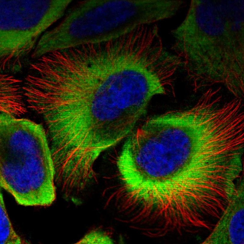
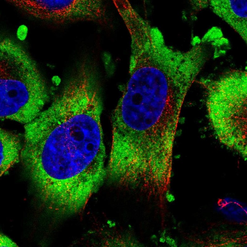
IMMUNOCYTOCHEMISTRY
Formal validation: Genetic
Relative fluorescence intensity (RFI)
Figure description
Antibody dilution
Standard validation
Literature conformity
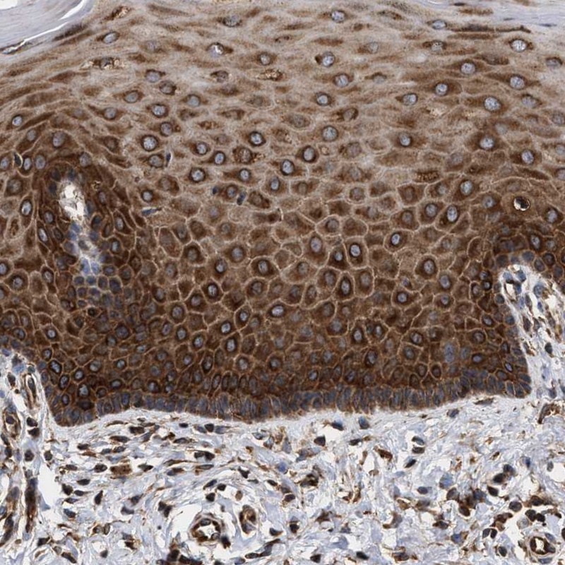
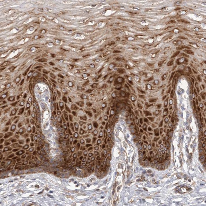
IMMUNOHISTOCHEMISTRY
Expression
Retrieval
RNA consistency
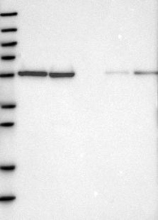
WESTERN BLOT
Target mass (kDa)
PROTEIN ARRAY
ANTIGEN INFORMATION
Antigen
Length (aa)
Antigen sequence
GHLQEGFGCVVTNRFDQLFDDESDPFEVLKAAENKKKEAGGGGVGGPGAK
SAAQAAAQTNSNAAGKQLRKESQKDRKNPLPPSVGVVDKKEETQPPVALK
KEGIRRVGRRPDQQLQGEGKIIDRRPERRPPRERRFE
Matching transcripts
RELEVANT PUBLICATIONS
1.
2.