We use cookies to enhance the usability of our website. If you continue, we'll assume that you are happy to receive all cookies. More information. Don't show this again.
Fields »
 

Field

Term
Gene name

Class

Subclass

Chromosome

External id

In atlas

Reliability

Reliability

Reliability

Validation

Method

Validation

Method

Validation

Method

Validation

Location

Searches

Tissue

Cell type

Expression

Tissue

Specificity

Cell line

Specificity

Tissue

Detectable

Cell line

Detectable

Category

Score

Score

Score

Antibodies

Column


ABOUT
HELP
BLOG
  • THE HUMAN PROTEOME
  • THE HUMAN TISSUES
  • THE HUMAN CELL
  • PROTEIN CLASSES
  • PROTEIN EVIDENCE
    • NEWS
    • BLOG
    • EVENTS
    • PRESS ROOM
    • LEARN
    • DICTIONARY
    • METHODS
    • CELL LINES
  • THE PROJECT
  • INTRODUCTION
  • ORGANIZATION
  • PUBLICATIONS
  • PUBLICATION DATA
  • ANTIBODY SUBMISSION
  • ANTIBODY AVAILABILITY
  • LINKS
  • CONTACT
  • TECHNICAL DATA
  • ANTIBODY VALIDATION
  • ASSAYS & ANNOTATION
  • DISCLAIMER
  • DOWNLOADABLE DATA
  • HELP & FAQ
  • LICENCE & CITATION
  • PRIVACY STATEMENT
  • RELEASE HISTORY
PDIA3
  • premium

    TISSUE

  • premium

    CELL

  • CANCER

ANTIBODY INFORMATION VALIDATION SUMMARY IMMUNOCYTOCHEMISTRY IMMUNOHISTOCHEMISTRY WESTERN BLOT PROTEIN ARRAY ANTIGEN INFORMATION RELEVANT PUBLICATIONS

ANTIBODY INFORMATION


Antibody HPA002645


Antibody HPA003230


Antibody CAB011199


Antibody CAB015181


Provider

Atlas Antibodies
Sigma-Aldrich
  Atlas Antibodies
Sigma-Aldrich
  SIGMA
  Santa Cruz Biotechnology
 

Product name

HPA002645   HPA003230   E5031   sc-28823  

Host species

Rabbit   Rabbit   Mouse   Rabbit  

Clonality

pAb   pAb   mAb   pAb  

Purity

Affinity purified using the PrEST-antigen as affinity ligand   Affinity purified using the PrEST-antigen as affinity ligand   Not known   Protein A/G  

Other gene match


Released in version

2   2   4   4  

References

1   4      

VALIDATION SUMMARY


Antibody HPA002645


Antibody HPA003230


Antibody CAB011199


Antibody CAB015181

 
Immunocytochemistry


Validated
 

Validated

Validated
     
Immunohistochemistry


Validated
 

Validated
  Supported   Supported  
Western blot

Supported  

Validated
  Supported   Supported  
Protein array

Supported   Supported      

IMMUNOCYTOCHEMISTRY


Antibody HPA002645


Antibody HPA003230


Antibody CAB011199


Antibody CAB015181


Formal validation: Genetic

 
Validated

Significant downregulation > 25% by both siRNA:s.


Relative fluorescence intensity (RFI)


Figure description

  Whole cells of segmented cells in 10x-images      

Antibody dilution

  1:127      
 

Formal validation: Independent

Validated

Antibody staining overlaps with antibody HPA003230.
 
Validated

Antibody staining overlaps with antibody HPA002645.


Standard validation

Supported   Supported      


Figure description

Immunofluorescent staining of human cell line U-2 OS shows localization to endoplasmic reticulum.   Immunofluorescent staining of human cell line U-2 OS shows localization to endoplasmic reticulum.      

Antibody dilution

1:20   1:125      

Literature conformity

The subcellular location is supported by literature.   The subcellular location is supported by literature.      

IMMUNOHISTOCHEMISTRY


Antibody HPA002645


Antibody HPA003230


Antibody CAB011199


Antibody CAB015181


Formal validation: Independent

Validated

Pearson correlation >0.6 for protein expression in cell lines using independent antibodies.
 
Validated

Pearson correlation >0.6 for protein expression in cell lines using independent antibodies.


Figure description

Distribution of protein expression (antibody staining).
Pearson correlation with HPA003230 across 46 cell lines.
  Distribution of protein expression (antibody staining).
Pearson correlation with HPA002645 across 46 cell lines.


Standard validation

Supported   Supported   Supported   Supported  


Figure description

Immunohistochemical staining of human thyroid gland shows strong cytoplasmic positivity in glandular cells.   Immunohistochemical staining of human thyroid gland shows strong cytoplasmic positivity in glandular cells.   Immunohistochemical staining of human thyroid gland shows strong cytoplasmic positivity in glandular cells.   Immunohistochemical staining of human thyroid gland shows strong cytoplasmic positivity in glandular cells.  

Expression

RNA: detected in 37 tissues
Protein: detected in 74 cell types
  RNA: detected in 37 tissues
Protein: detected in 63 cell types
  RNA: detected in 37 tissues
Protein: detected in 65 cell types
  RNA: detected in 37 tissues
Protein: detected in 62 cell types
 

Retrieval

HIER pH6   HIER pH6   HIER pH6   HIER pH6  

Antibody dilution

1:500   1:6000   1:200   1:400  

Literature conformity

Consistent with extensive gene/protein characterization data.   Consistent with extensive gene/protein characterization data.   Consistent with extensive gene/protein characterization data.   Consistent with extensive gene/protein characterization data.  

RNA consistency

Consistent with RNA expression data.   Consistent with RNA expression data.   Consistent with RNA expression data.   Consistent with RNA expression data.  

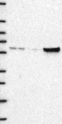
WESTERN BLOT


Antibody HPA002645


Antibody HPA003230


Antibody CAB011199


Antibody CAB015181


Formal validation: Genetic

 
Validated

Downregulation visible in one of two siRNA lanes


Figure description

  Lane 1: Marker [kDa] 250, 130, 95, 72, 55, 36, 28, 17, 10
Lane 2: siRNA 1
Lane 3: siRNA 2
Lane 4: Scrambled
     

Target mass (kDa)

  56.8      

Loading control

  Total protein image      

Antibody dilution

  1:281      
 

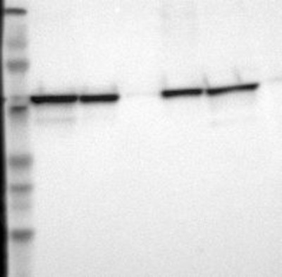
Standard validation

Supported

Analysis performed using a standard panel of samples.
Band of predicted size in kDa (+/-20%) with additional bands present.
  Supported

Analysis performed using a standard panel of samples.
Single band corresponding to the predicted size in kDa (+/-20%).
  Supported

Analysis performed using a standard panel of samples.
Band of predicted size in kDa (+/-20%) with additional bands present.
  Supported

Analysis performed using a standard panel of samples.
Band of predicted size in kDa (+/-20%) with additional bands present.


Figure description

Lane 1: Marker [kDa] 220, 112, 84, 47, 32, 26, 16.8
Lane 2: RT4
Lane 3: U-251 MG
Lane 4: Liver
Lane 5: Tonsil
Lane 6:
  Lane 1: Marker [kDa] 220, 112, 84, 47, 32, 26, 16.8
Lane 2: RT4
Lane 3: U-251 MG
Lane 4: Human Plasma
Lane 5: Liver
Lane 6: Tonsil
  Lane 1: Marker [kDa] 219, 111, 83, 48, 32, 26, 17
Lane 2: RT4
Lane 3: U-251 MG
Lane 4: Human Plasma
Lane 5: Liver
Lane 6: Tonsil
  Lane 1: Marker [kDa] 229, 107, 78, 46, 31, 26, 16.2
Lane 2: RT4
Lane 3: U-251 MG
Lane 4: Human Plasma
Lane 5: Liver
Lane 6: Tonsil
 

Target mass (kDa)

56.8   56.8   56.8   56.8  

Antibody dilution

1:250   1:250   1:500   1:500  

PROTEIN ARRAY


Antibody HPA002645


Antibody HPA003230


Antibody CAB011199


Antibody CAB015181


Standard validation

Supported

Pass with single peak corresponding to interaction only with its own antigen.
  Supported

Pass with single peak corresponding to interaction only with its own antigen.


Figure description

Antibody specificity analysis with protein arrays. Predicted and matching interactions are shown in green.   Antibody specificity analysis with protein arrays. Predicted and matching interactions are shown in green.      

Antibody dilution

1:2000   1:3000      

ANTIGEN INFORMATION


Antibody HPA002645


Antibody HPA003230


Antibody CAB011199


Antibody CAB015181


Antigen

Recombinant protein fragment   Recombinant protein fragment   Native protein   Recombinant protein  

Length (aa)

132   118      

Antigen sequence

HMTEDNKDLIQGKDLLIAYYDVDYEKNAKGSNYWRNRVMMVAKKFLDAGH
KLNFAVASRKTFSHELSDFGLESTAGEIPVVAIRTAKGEKFVMQEEFSRD
GKALERFLQDYFDGNLKRYLKSEPIPESNDGP
 
PTLKIFRDGEEAGAYDGPRTADGIVSHLKKQAGPASVPLRTEEEFKKFIS
DKDASIVGFFDDSFSEAHSEFLKAASNLRDNYRFAHTNVESLVNEYDDNG
EGIILFRPSHLTNKFEDK
     

Matching transcripts

PDIA3-001 - ENSP00000300289 [100%]
  PDIA3-001 - ENSP00000300289 [100%]
     

Other gene match

       
ANTIGEN VIEW
PDIA3-001

RELEVANT PUBLICATIONS


Antibody HPA002645


Antibody HPA003230


Antibody CAB011199


Antibody CAB015181


1.

SILAC-based quantitative proteomic approach to identify potential biomarkers from the esophageal squamous cell carcinoma secretome
Kashyap MK et al
Cancer Biol Ther 2010;10(8):796-810
  The involvement of SMILE/TMTC3 in endoplasmic reticulum stress response
Racapé M et al
PLoS One 2011;6(5):e19321
     

2.

  Loss-of-function mutations in MICU1 cause a brain and muscle disorder linked to primary alterations in mitochondrial calcium signaling
Logan CV et al
Nat Genet 2014;46(2):188-93
     

3.

  Proteomics based identification of cell migration related proteins in HBV expressing HepG2 cells
Feng H et al
PLoS One 2014;9(4):e95621
Application: WB
     

4.

  The dehydrogenase region of the NADPH oxidase component Nox2 acts as a protein disulfide isomerase (PDI) resembling PDIA3 with a role in the binding of the activator protein p67 (phox.)
Bechor E et al
Front Chem 2015;3:3
Application: WB


Contact

  • BLOG
  • PRESS ROOM
  • contact@proteinatlas.org

The Project

  • INTRODUCTION
  • ORGANIZATION
  • PUBLICATIONS
  • DOWNLOADABLE DATA

The Human Protein Atlas

  • ANTIBODY AVAILABILITY
  • LICENCE & CITATION
  • HELP & FAQ
The Human Protein Atlas project is funded
by the Knut & Alice Wallenberg foundation.