We use cookies to enhance the usability of our website. If you continue, we'll assume that you are happy to receive all cookies. More information. Don't show this again.
Fields »
 

Field

Term
Gene name

Class

Subclass

Chromosome

External id

In atlas

Reliability

Reliability

Reliability

Validation

Method

Validation

Method

Validation

Method

Validation

Location

Searches

Tissue

Cell type

Expression

Tissue

Specificity

Cell line

Specificity

Tissue

Detectable

Cell line

Detectable

Category

Score

Score

Score

Antibodies

Column


ABOUT
HELP
BLOG
  • THE HUMAN PROTEOME
  • THE HUMAN TISSUES
  • THE HUMAN CELL
  • PROTEIN CLASSES
  • PROTEIN EVIDENCE
    • NEWS
    • BLOG
    • EVENTS
    • PRESS ROOM
    • LEARN
    • DICTIONARY
    • METHODS
    • CELL LINES
  • THE PROJECT
  • INTRODUCTION
  • ORGANIZATION
  • PUBLICATIONS
  • PUBLICATION DATA
  • ANTIBODY SUBMISSION
  • ANTIBODY AVAILABILITY
  • LINKS
  • CONTACT
  • TECHNICAL DATA
  • ANTIBODY VALIDATION
  • ASSAYS & ANNOTATION
  • DISCLAIMER
  • DOWNLOADABLE DATA
  • HELP & FAQ
  • LICENCE & CITATION
  • PRIVACY STATEMENT
  • RELEASE HISTORY
S1PR1
  • premium

    TISSUE

  • premium

    CELL

  • CANCER

CANCER ATLAS

GENE/PROTEIN

Antibody validation

Dictionary

Level of antibody staining/expression


High
Medium
Low
Not detected

CANCER ATLASi

The cancer tissue page shows antibody staining in 20 different cancers. The assay and annotation is described here.

This page starts with information about the protein evidence and, when applicable, also protein class. Below the summary, a selection of four standard cancer tissue samples is displayed as representative of the overall staining pattern. From left: colorectal cancer, breast cancer, prostate cancer and lung cancer. An additional 5th image can be added as a complement.

For histological reference, visit the histological dictionary.

Gene description

Sphingosine-1-phosphate receptor 1

Protein class

Cancer-related genes, CD markers, G-protein coupled receptors, Predicted membrane proteins, Transporters

Protein evidence

Evidence at protein level
Colorectal cancer
Breast cancer
Prostate cancer
Lung cancer
Glioma

STAINING SUMMARYi

The cancer tissue summary page shows antibody staining in 20 different cancers. The assay and annotation is described here.

For each cancer, the fraction of samples with antibody staining/protein expression level high, medium, low, or not detected are provided by the blue-scale color-coding (as described by the color-coding scale in the box to the right). The length of the bar represents the number of patient samples analyzed (max=12 patients). The images and annotations can be accessed by clicking on the cancer name or protein expression bar. If more than one antibody is analyzed, the tabs at the top of the staining summary section can be used to toggle between the different antibodies. The tooltip function displays additional data for the features in the staining summary view.

Next to the cancer staining data, the protein expression data of normal tissues or specific cell types corresponding to each cancer are shown and protein expression levels are indicated by the blue-scale color coding.

At the bottom of the page, a summary of the overall protein expression pattern across the analyzed cancer tissues as well as information about literature conformity are presented.

CAB010104

Tissue

Cancer staining

Protein
expression of
normal tissue

Breast cancer
 
Carcinoid
 
Cervical cancer
 
 
Colorectal cancer
 
 
Endometrial cancer
 
 
Glioma
 
Head and neck cancer
 
 
Liver cancer
 
 
Lung cancer
 
 
Lymphoma


Tissue

Cancer staining

Protein
expression of
normal tissue

Melanoma
 
Ovarian cancer
 
Pancreatic cancer
 
Prostate cancer
 
Renal cancer
 
Skin cancer
 
Stomach cancer
 
 
Testis cancer
 
Thyroid cancer
 
Urothelial cancer
 

Staining summary

Weak to moderate cytoplasmic staining was observed in most malignancies. Several endometrial and thyroid cancers as well as malignant gliomas were strongly stained.
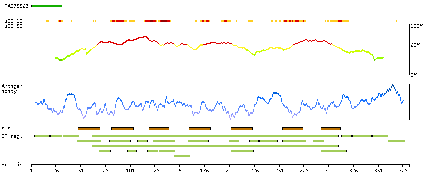
GENE INFORMATIONi

Gene information from Ensembl and Entrez, as well as links to available gene identifiers are displayed here. Information was retrieved from Ensembl if not indicated otherwise.

Gene name

S1PR1 (HGNC Symbol)

Synonyms

CD363, D1S3362, edg-1, EDG1

Description

Sphingosine-1-phosphate receptor 1 (HGNC Symbol)

Entrez gene summary

The protein encoded by this gene is structurally similar to G protein-coupled receptors and is highly expressed in endothelial cells. It binds the ligand sphingosine-1-phosphate with high affinity and high specificity, and suggested to be involved in the processes that regulate the differentiation of endothelial cells. Activation of this receptor induces cell-cell adhesion. [provided by RefSeq, Jul 2008]

Chromosome

1

Cytoband

p21.2

Chromosome location (bp)

101236888 - 101241518

Protein evidence

Evidence at protein level (all genes)

Ensembl

ENSG00000170989 (version 83.38)

Entrez gene

1901

UniProt

P21453 (UniProt - Evidence at protein level)

neXtProt

NX_P21453

Antibodypedia

S1PR1 antibodies

PROTEIN BROWSERi

The protein browser displays the antigen location on the target protein(s) and the features of the target protein. The tabs at the top of the protein view section can be used to switch between the different splice variants to which an antigen has been mapped.

At the top of the view, the position of the antigen (identified by the corresponding HPA identifier) is shown as a green bar. A yellow triangle on the bar indicates a <100% sequence identity to the protein target.

Under the antigens, the maximum percent sequence identity of the protein to all other proteins from other human genes is displayed, using a sliding window of 10 aa residues (HsID 10) or 50 aa residues (HsID 50) (read more).

If a signal peptide is predicted by a majority of the signal peptide predictors SPOCTOPUS, SignalP 4.0, and Phobius (turquoise) and/or transmembrane regions (orange) are predicted by MDM, these are displayed.

Low complexity regions are shown in yellow and InterPro regions in green. Common (purple) and unique (grey) regions between different splice variants of the gene are also displayed (read more), and at the bottom of the protein view is the protein scale.

S1PR1-001

PROTEIN INFORMATIONi

The protein information section displays alternative protein-coding transcripts (splice variants) encoded by this gene according to the Ensembl database.

The ENSP identifier links to the Ensembl website protein summary, while the ENST identifier links to the Ensembl website transcript summary for the selected splice variant. The data in the UniProt column can be expanded to show links to all matching UniProt identifiers for this protein.

The protein classes assigned to this protein are shown if expanding the data in the protein class column. Parent protein classes are in bold font and subclasses are listed under the parent class.

The Gene Ontology terms assigned to this protein are listed if expanding the Gene ontology column. The length of the protein (amino acid residues according to Ensembl), molecular mass (kDalton), predicted signal peptide (according to a majority of the signal peptide predictors SPOCTOPUS, SignalP 4.0, and Phobius) and the number of predicted transmembrane region(s) (according to MDM) are also reported.

Splice variant

UniProt

Protein class

Gene ontology

Length & mass

Signal peptide
(predicted)

Transmembrane regions
(predicted)

S1PR1-001
ENSP00000305416
ENST00000305352
P21453 [Direct mapping]
Sphingosine 1-phosphate receptor 1
Show all
CD markers
Transporters
G-protein coupled receptors
   GPCRs excl olfactory receptors
   Lysolipids receptors
Predicted membrane proteins
   Prediction method-based
   Membrane proteins predicted by MDM
   MEMSAT3 predicted membrane proteins
   MEMSAT-SVM predicted membrane proteins
   Phobius predicted membrane proteins
   SCAMPI predicted membrane proteins
   SPOCTOPUS predicted membrane proteins
   THUMBUP predicted membrane proteins
   TMHMM predicted membrane proteins
   GPCRHMM predicted membrane proteins
   # TM segments-based
   7TM proteins predicted by MDM
Cancer-related genes
   Candidate cancer biomarkers
Protein evidence (Kim et al 2014)
Show all
GO:0001525 [angiogenesis]
GO:0001664 [G-protein coupled receptor binding]
GO:0001955 [blood vessel maturation]
GO:0003245 [cardiac muscle tissue growth involved in heart morphogenesis]
GO:0003376 [sphingosine-1-phosphate signaling pathway]
GO:0004930 [G-protein coupled receptor activity]
GO:0005768 [endosome]
GO:0005829 [cytosol]
GO:0005886 [plasma membrane]
GO:0006935 [chemotaxis]
GO:0007155 [cell adhesion]
GO:0007186 [G-protein coupled receptor signaling pathway]
GO:0007193 [adenylate cyclase-inhibiting G-protein coupled receptor signaling pathway]
GO:0007420 [brain development]
GO:0008284 [positive regulation of cell proliferation]
GO:0009897 [external side of plasma membrane]
GO:0016021 [integral component of membrane]
GO:0016477 [cell migration]
GO:0019226 [transmission of nerve impulse]
GO:0030032 [lamellipodium assembly]
GO:0030155 [regulation of cell adhesion]
GO:0030182 [neuron differentiation]
GO:0030335 [positive regulation of cell migration]
GO:0030500 [regulation of bone mineralization]
GO:0031226 [intrinsic component of plasma membrane]
GO:0031532 [actin cytoskeleton reorganization]
GO:0038036 [sphingosine-1-phosphate receptor activity]
GO:0043547 [positive regulation of GTPase activity]
GO:0045121 [membrane raft]
GO:0045124 [regulation of bone resorption]
GO:0045446 [endothelial cell differentiation]
GO:0045944 [positive regulation of transcription from RNA polymerase II promoter]
GO:0046625 [sphingolipid binding]
GO:0048661 [positive regulation of smooth muscle cell proliferation]
GO:0050927 [positive regulation of positive chemotaxis]
GO:0051482 [positive regulation of cytosolic calcium ion concentration involved in phospholipase C-activating G-protein coupled signaling pathway]
GO:0051497 [negative regulation of stress fiber assembly]
GO:0061384 [heart trabecula morphogenesis]
GO:0072678 [T cell migration]
Show all
382 aa
42.8 kDa
No 7

Contact

  • BLOG
  • PRESS ROOM
  • contact@proteinatlas.org

The Project

  • INTRODUCTION
  • ORGANIZATION
  • PUBLICATIONS
  • DOWNLOADABLE DATA

The Human Protein Atlas

  • ANTIBODY AVAILABILITY
  • LICENCE & CITATION
  • HELP & FAQ
The Human Protein Atlas project is funded
by the Knut & Alice Wallenberg foundation.