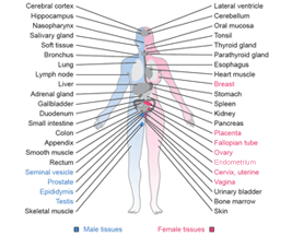
Liver
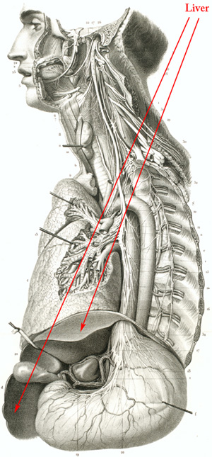
Liver
The classical hexagonal liver lobule is surrounded on an average by six portal tracts and centered on a terminal twig of the hepatic vein: the centrolobular or central vein. The portal vein ramifications in the portal tracts give off a series of branches in the plane between adjacent portal tracts; these give rise to the sinusoids that drain the blood towards the centre of the lobule. The lobule may be viewed to comprise a periportal zone, midzonal area and centrilobular area.
The hepatocyte is a polygonal cell that usually contains a single, central nucleus and occasional brownish pigment representing intracellular bile. The hepatocytes are usually arranged in one-cell thick plates called muralia with a sinusoid on either side thus exposing the hepatocyte to portal blood on two surfaces. Within the muralium each hepatocyte adjoins the adjacent cell with its intercellular surface. The intercellular domain of the cell membrane carries a groove termed the hemicanaliculus. The hemicanaliculus of two adjacent hepatocytes comprises the intercellular bile canaliculus.
The space of Disse is formed between the sinusoidal lining cells and the sinusoidal domain of the hepatocyte surface. Several different cell types including sinusoidal endothelial cells, Kupffer cells, hepatic stellate cells (Ito cells) line the hepatic sinusoids, each having its own special function.
The portal tracts at the lobular periphery are composed of connective tissue ensheathing branches of the hepatic artery, portal vein, bile duct (together termed the portal triad) and lymphatics. The caliber of the portal tracts decreases from the hilum of the liver towards its periphery.
Cancer: Liver cancer
|