TISSUE
CELL
CANCER
ANTIBODY INFORMATION
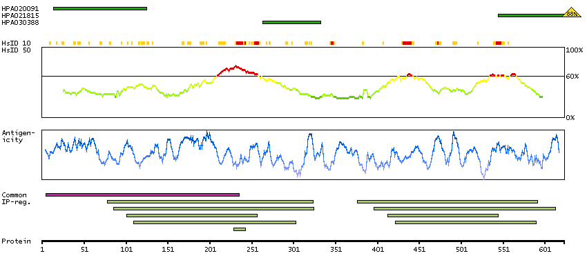
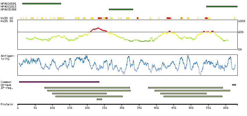
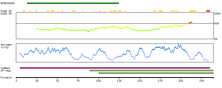
Antibody HPA020091
Antibody HPA021815
Antibody HPA030388
Antibody CAB020682
Provider
Product name
Host species
Clonality
Purity
Other gene match
Released in version
References
VALIDATION SUMMARY
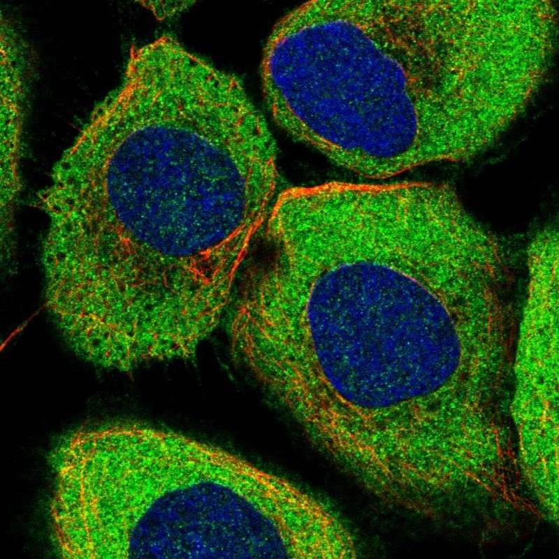
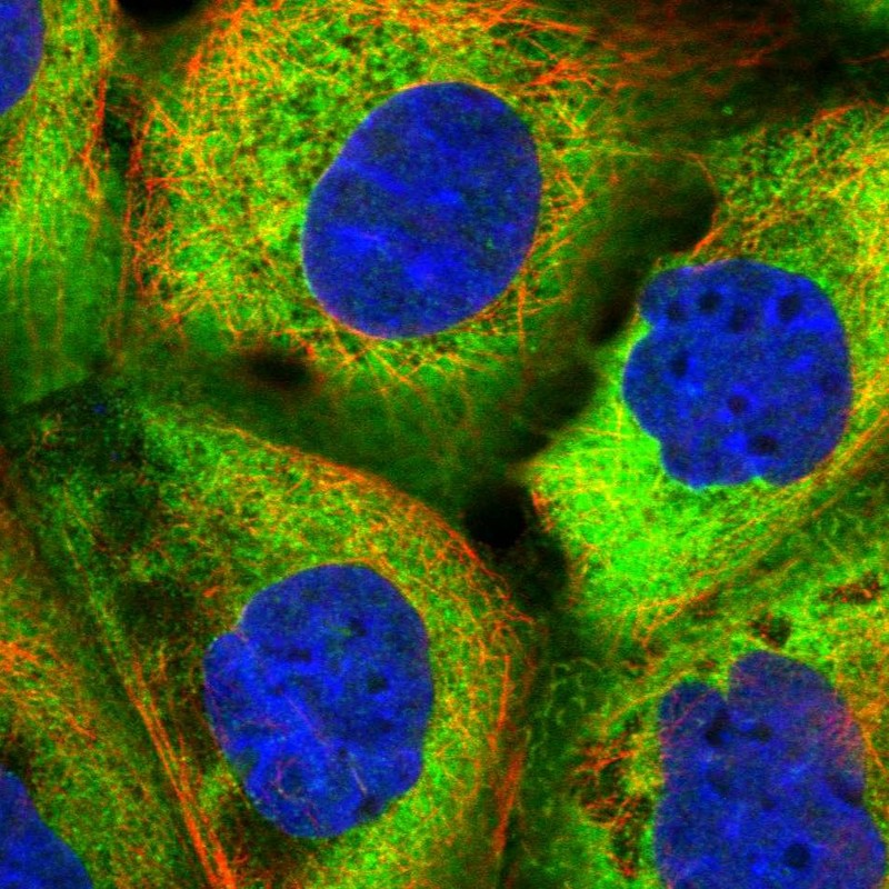
IMMUNOCYTOCHEMISTRY
Standard validation
Figure description
Antibody dilution
Literature conformity
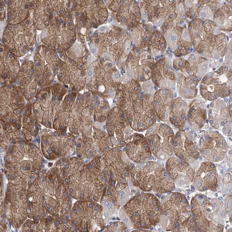
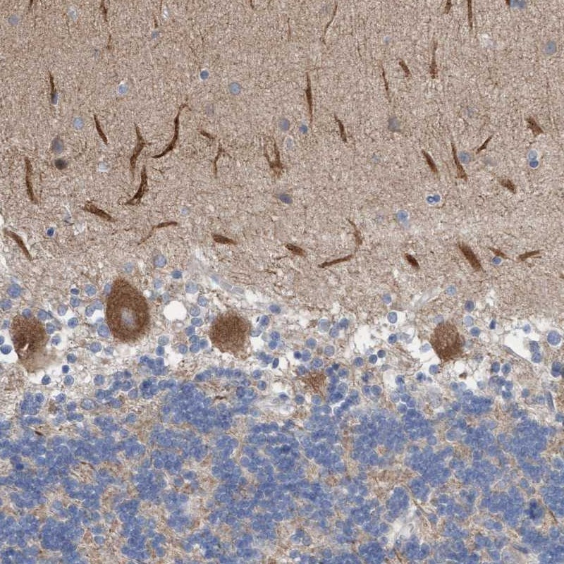
IMMUNOHISTOCHEMISTRY
Expression
Retrieval
RNA consistency
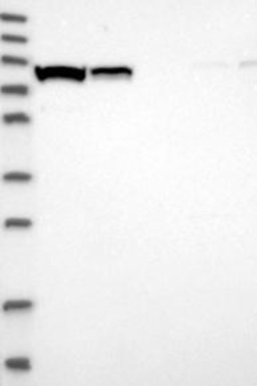
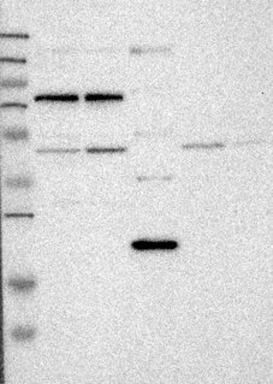
WESTERN BLOT
Target mass (kDa)
PROTEIN ARRAY
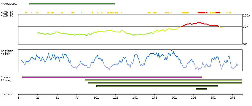
ANTIGEN INFORMATION
Antigen
Length (aa)
Antigen sequence
KEAAKARQRPRKGHEENGDVVTEPQVAEKNEANGRETTEVDLLTKELEDF
EMKKAAARAVTGVLASHPNSTDVHIINLSLTFHGQELLSDTKLELNSGRR
YGLIGLNGIGKS
NHLDIETIDALADAINEFEGGMMLVSHDFRLIQQVAQEIWVCEKQTITKW
PGDILAYKEHLKSKLVDEEPQLTKRTHNVCTLTLASLPRP
CVWLEEELKTFKRILVLVSHSQDFLNGVCTNIIHMHNKKLKYYTGNYDQY
VKTRLELEENQMKRFHWEQD
Matching transcripts