TISSUE
CELL
CANCER
ANTIBODY INFORMATION
Antibody HPA002828
Antibody HPA002829
Antibody CAB016446
Provider
Product name
Host species
Clonality
Purity
Other gene match
Released in version
References
VALIDATION SUMMARY
IMMUNOCYTOCHEMISTRY
Standard validation
Figure description
Antibody dilution
Literature conformity
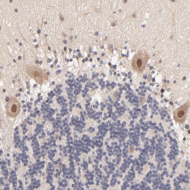
IMMUNOHISTOCHEMISTRY
Formal validation: Independent
Expression
Retrieval
RNA consistency
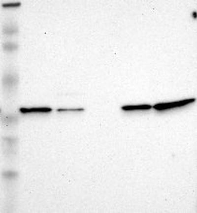
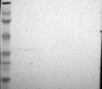
WESTERN BLOT
Target mass (kDa)
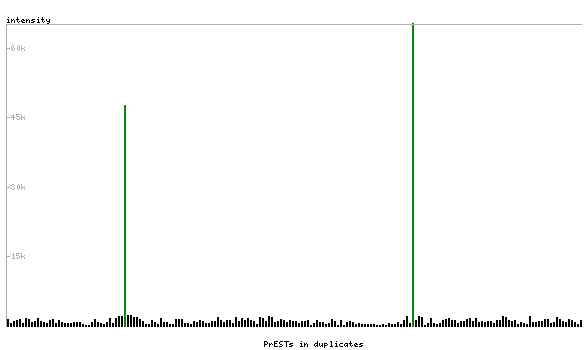
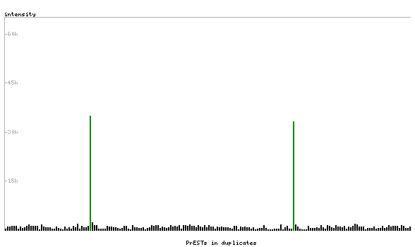
PROTEIN ARRAY
ANTIGEN INFORMATION
Antigen
Length (aa)
Antigen sequence
CLNESDEHGFDNCLRKDTTFLESDCDEQLLITVAFNQPVKLYSMKFQGPD
NGQGPKYVKIFINLPRSMDFEEAERSEPTQALELTEDDIKEDGIVPLRYV
KFQNVNSVTIFVQSNQGEEETTRISYFTFIGTPVQATNMNDFKRVVGK
LLLQFQSQWRRGHPAGLSVRMVGVKPVGSDPDFQPELSGAGSRLAVVKFT
MRGCGPCLRIAPAFSSMSNKYPQAVFLEVDVHQCQGTAATNNISATPTFL
FFRNKVRIDQYQGADAVGLEEKIKQHLENDPGSNEDTDIPKG
Matching transcripts