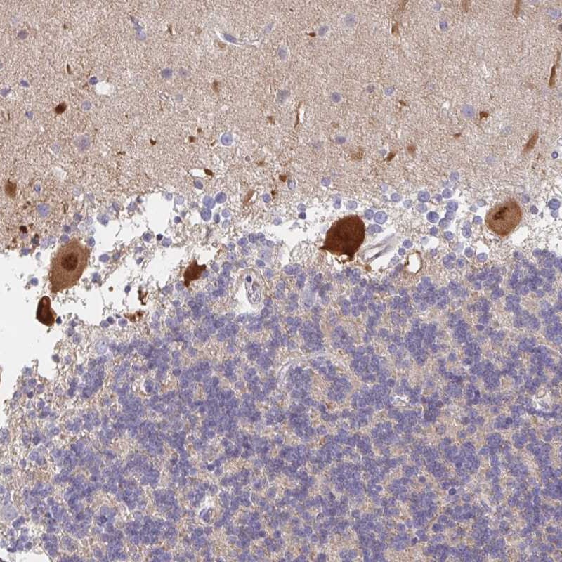
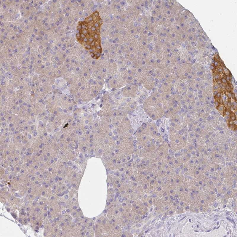
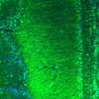
TISSUE
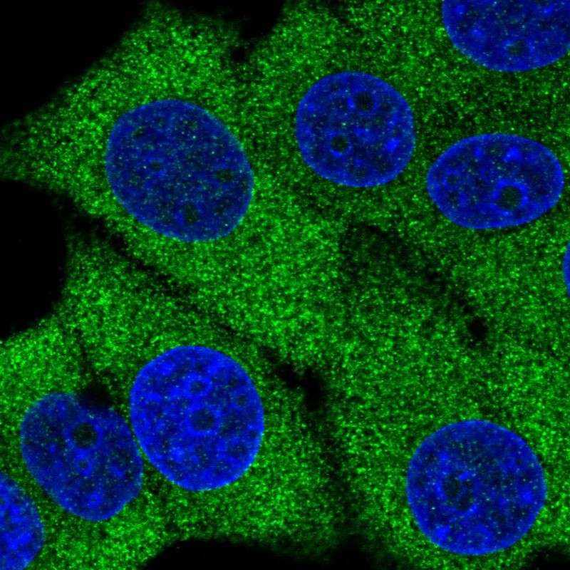
CELL
CANCER
ANTIBODY INFORMATION
Antibody HPA027137
Antibody HPA045760
Antibody HPA050590
Provider
Product name
Host species
Clonality
Purity
Other gene match
Released in version
References
VALIDATION SUMMARY
IMMUNOCYTOCHEMISTRY
Formal validation: Genetic
Relative fluorescence intensity (RFI)
Figure description
Antibody dilution
Standard validation
Literature conformity
IMMUNOHISTOCHEMISTRY
Formal validation: Independent
Expression
Retrieval
RNA consistency
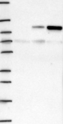
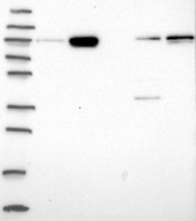
WESTERN BLOT
Target mass (kDa)
Loading control
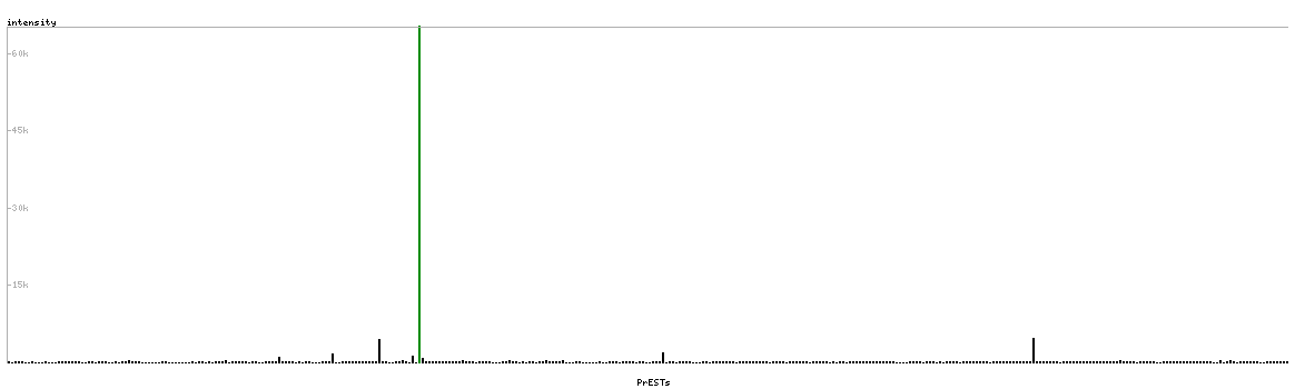
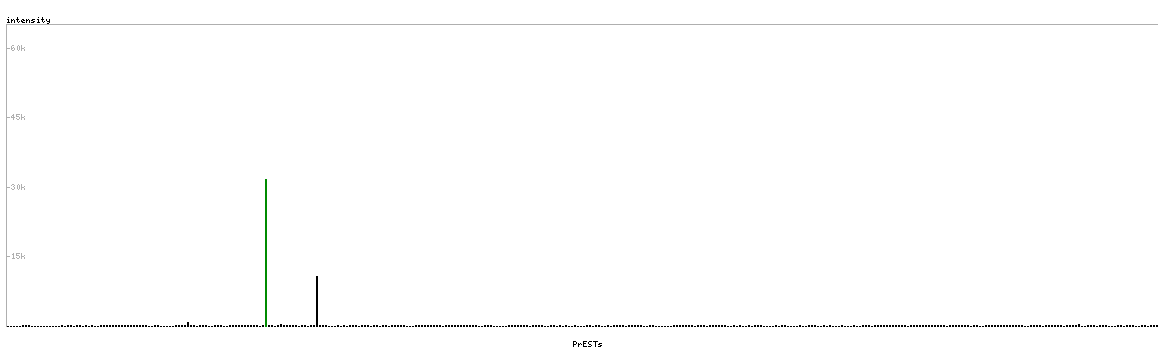
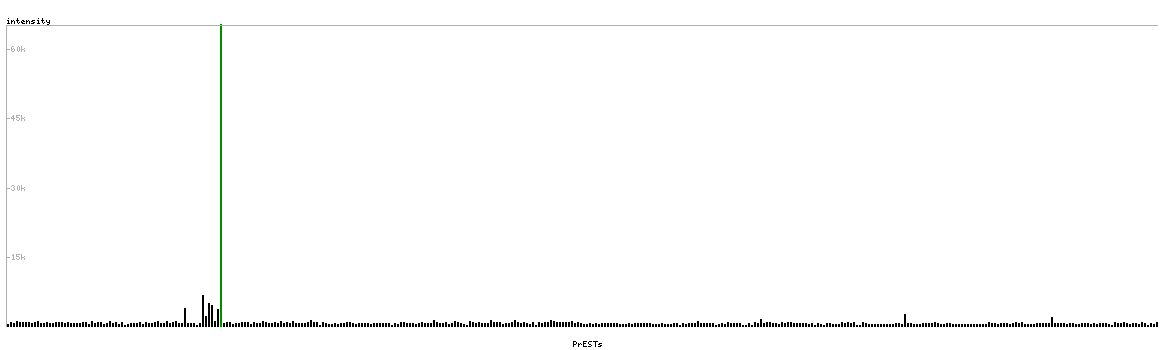
PROTEIN ARRAY
ANTIGEN INFORMATION
Antigen
Length (aa)
Antigen sequence
VLEREFQRVTISGEEKCGVPFTDLLDAAKSVVRALFIREKYMALSLQSFC
PTTRRYLQQLAEKPLETRTYEQGPDTPVSADAP
MDGKCKEIAEELFTRSLAESELRSAPYEFPEESPIEQLEERRQRLERQIS
QDVKLEPDILLRAKQDFLKTDSDSDLQLYKEQGE
RRKGLDVAEPGPSRCRSDSPAVAAVVPAMASYPSGSGKPKAKYPFKKRAS
LQASTAAPEARGGLGAPPLQSARSLPGPAPCLKHFPL
Matching transcripts