TISSUE
CELL
CANCER
ANTIBODY INFORMATION
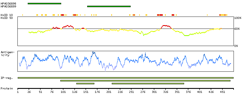
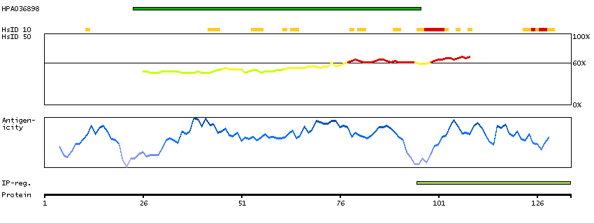
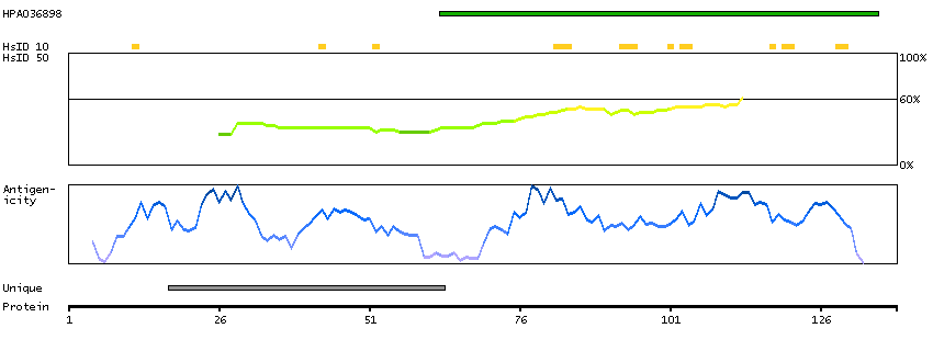
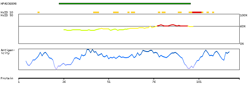
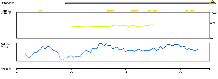
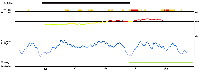
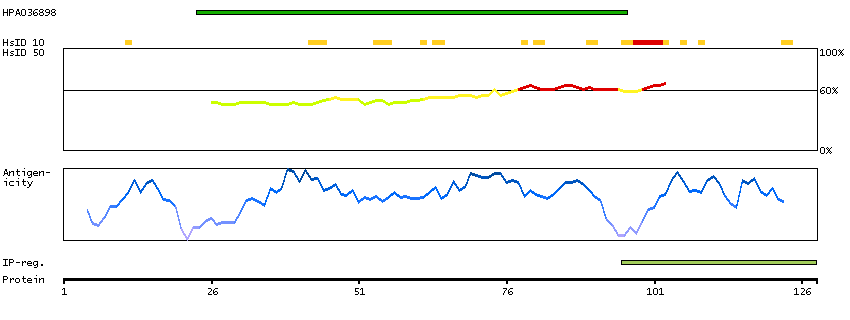
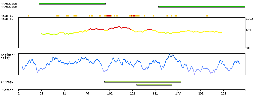
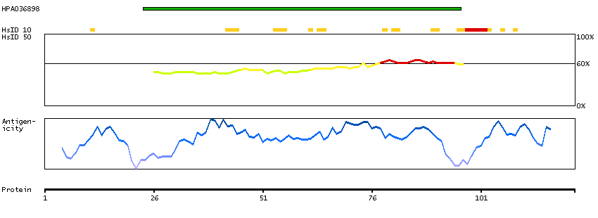
Antibody HPA036898
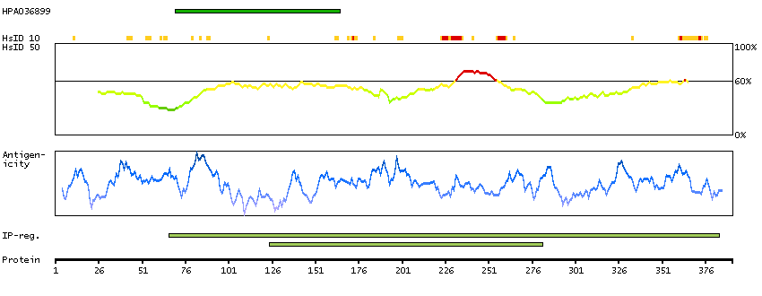
Antibody HPA036899
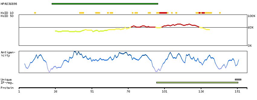
Antibody CAB026196
Provider
Product name
Host species
Clonality
Purity
Other gene match
Released in version
References
VALIDATION SUMMARY
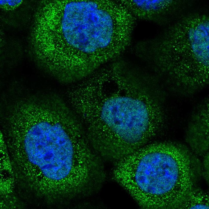
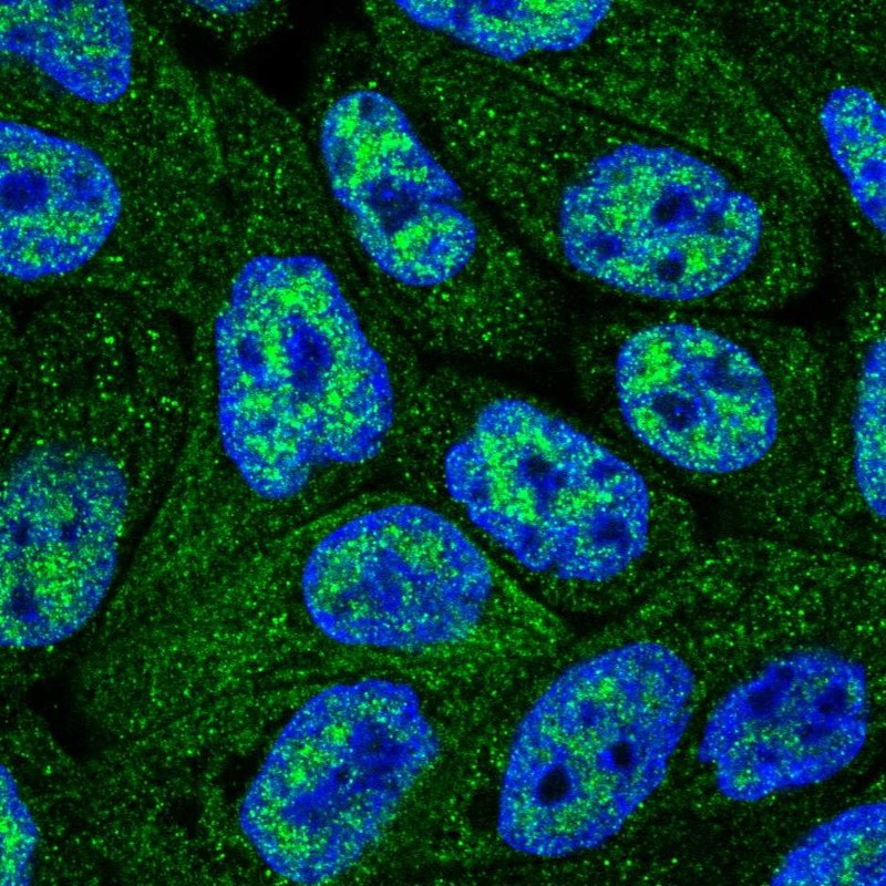
IMMUNOCYTOCHEMISTRY
Formal validation: Independent
Standard validation
Figure description
Antibody dilution
Literature conformity
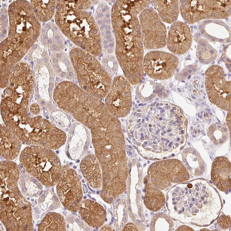
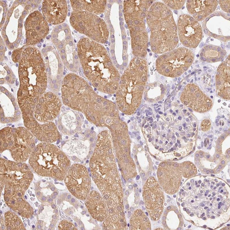
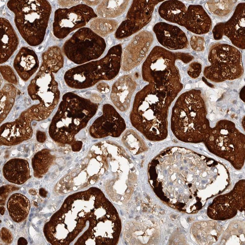
IMMUNOHISTOCHEMISTRY
Formal validation: Orthogonal
Expression
Retrieval
RNA consistency
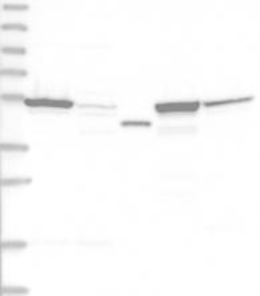
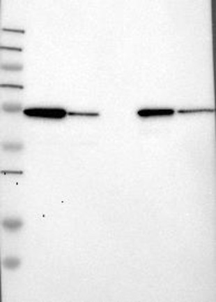
WESTERN BLOT
Target mass (kDa)
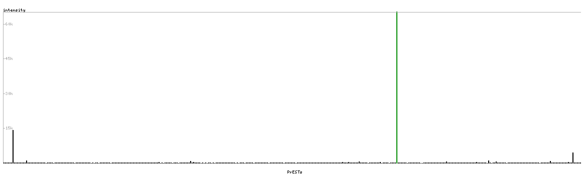
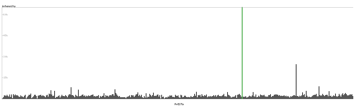
PROTEIN ARRAY
ANTIGEN INFORMATION
Antigen
Length (aa)
Antigen sequence
AKWVAIQSVSAWPEKRGEIRRMMEVAAADVKQLGGSVELVDIGKQKLPDG
SEIPLPPILLGRLGSDPQKKTVCI
IPVNVRFCLEGMEESGSEGLDELIFARKDTFFKDVDYVCISDNYWLGKKK
PCITYGLRGICYFFIEVECSNKDLHSGVYGGSVHEAMTDLILLMGS
Matching transcripts