TISSUE
CELL
CANCER
ANTIBODY INFORMATION
Antibody HPA045168
Antibody CAB000147
Antibody CAB003839
Antibody CAB003840
Antibody CAB075726
Antibody CAB075727
Provider
Product name
Host species
Clonality
Purity
Other gene match
Released in version
References
VALIDATION SUMMARY
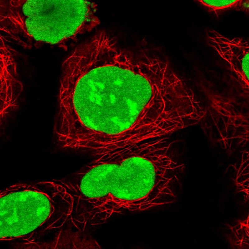
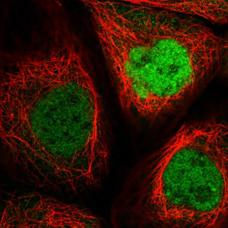
IMMUNOCYTOCHEMISTRY
Formal validation: Genetic
Relative fluorescence intensity (RFI)
Figure description
Antibody dilution
Standard validation
Literature conformity
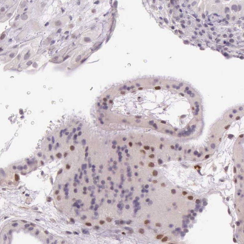
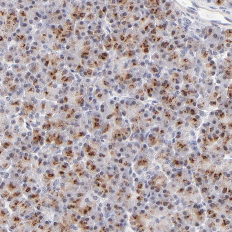
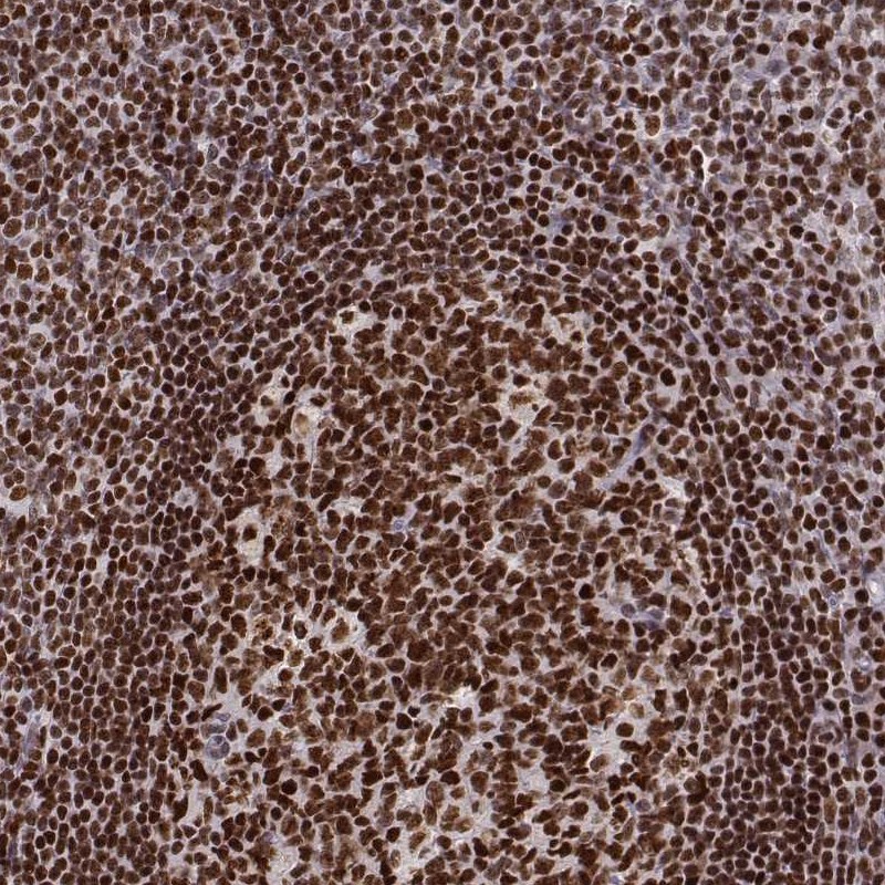
IMMUNOHISTOCHEMISTRY
Formal validation: Orthogonal
Expression
Retrieval
RNA consistency
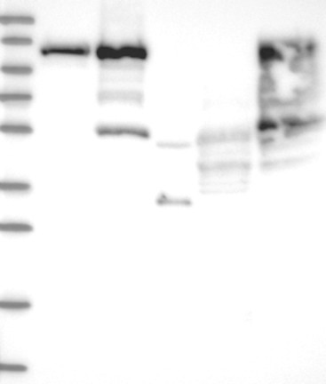
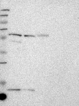
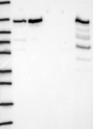
WESTERN BLOT
Target mass (kDa)
Loading control
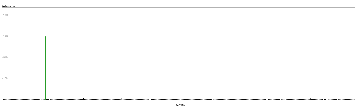
PROTEIN ARRAY
ANTIGEN INFORMATION
Antigen
Length (aa)
Antigen sequence
KGGKVFSATLGLVDIVKGTNSYYKLQLLEDDKENRYWIFRSWGRVGTVIG
SNKLEQMPSKEDAIEHFMKLYEEKTGNAWHSKNFTKYPKKFYPLEIDYGQ
DEEAVKKLTVNPGTKSKLPKPVQDLIKMIFDVESMKKAM
Matching transcripts