We use cookies to enhance the usability of our website. If you continue, we'll assume that you are happy to receive all cookies. More information. Don't show this again.
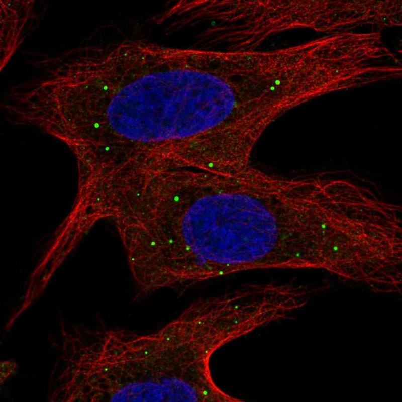
Immunofluorescent staining of human cell line U-2 OS shows localization to vesicles.
Antibody dilution
1:23
Literature conformity
The subcellular location is supported by literature.
IMMUNOHISTOCHEMISTRY
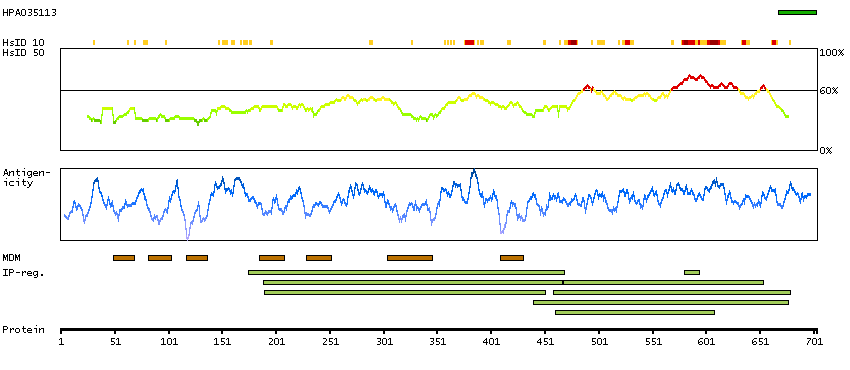
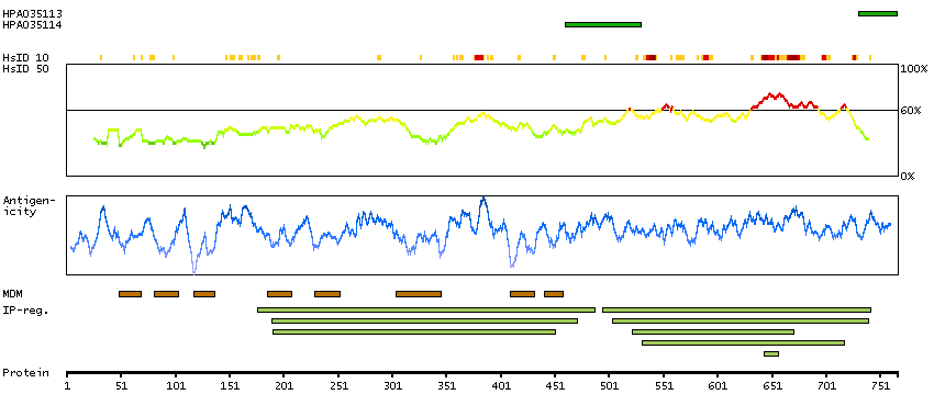
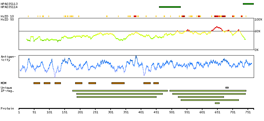
Antibody HPA035113
Antibody HPA035114
Antibody CAB033052
Standard validation
Approved
Approved
Figure description
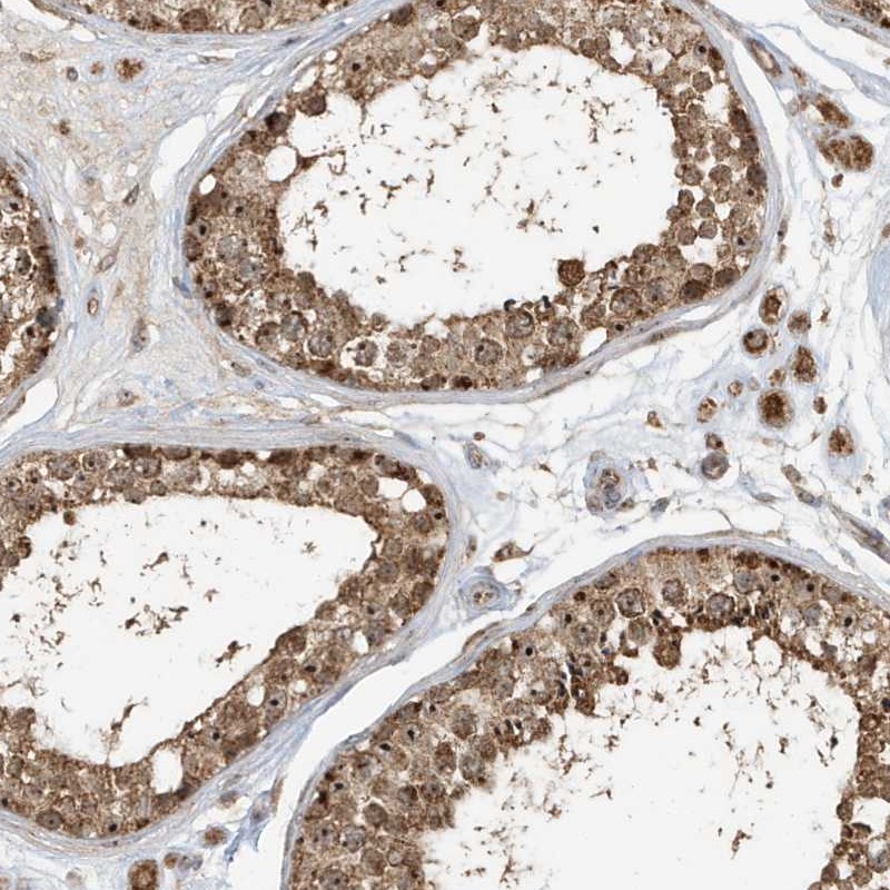
Immunohistochemical staining of human testis shows strong cytoplasmic positivity.
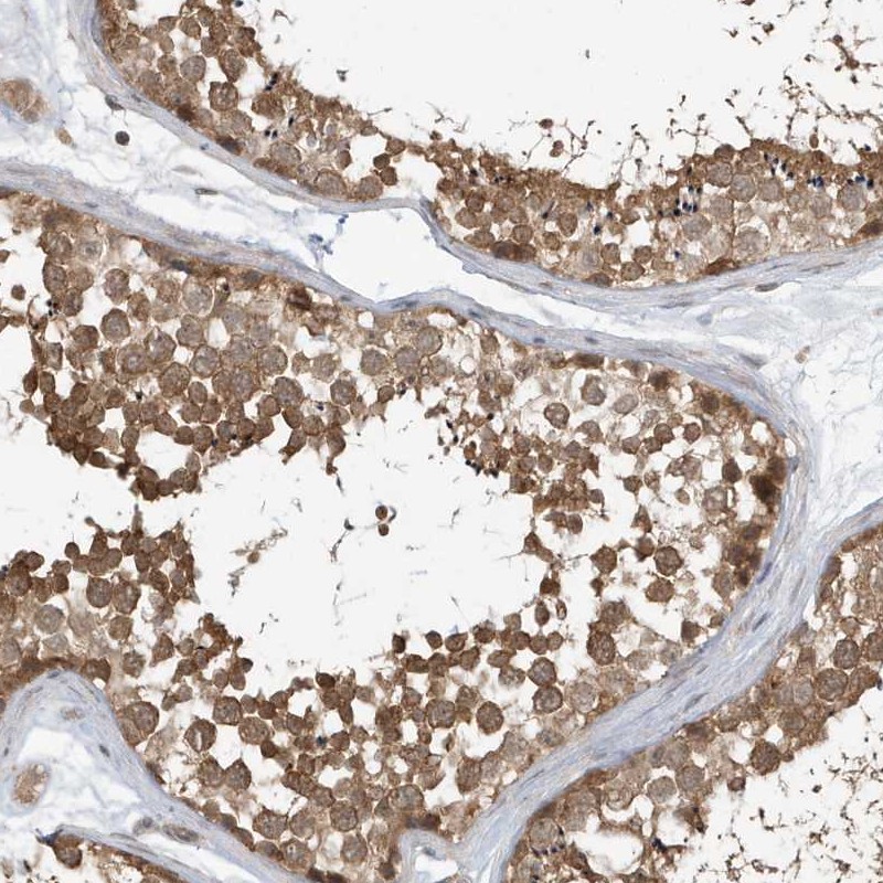
Immunohistochemical staining of human testis shows strong cytoplasmic positivity in cells in seminiferus ducts.
Expression
RNA: detected in 34 tissues Protein: detected in 76 cell types
RNA: detected in 34 tissues Protein: detected in 72 cell types
Retrieval
HIER pH6
HIER pH6
Antibody dilution
1:25
1:25
Literature conformity
Partly consistent with gene/protein characterization data.
Consistent with gene/protein characterization data.
RNA consistency
Mainly not consistent with RNA expression data.
Mainly not consistent with RNA expression data.
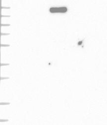
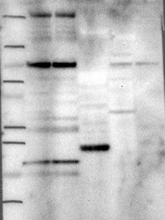
WESTERN BLOT
Antibody HPA035113
Antibody HPA035114
Antibody CAB033052
Standard validation
Uncertain
Analysis performed using a standard panel of samples. Single band larger than predicted size in kDa (+20%) but partly supported by experimental and/or bioinformatic data.
Uncertain
Analysis performed using a standard panel of samples. Weak band of predicted size but with additional bands of higher intensity also present.
Supported
Analysis performed using a standard panel of samples. Band of predicted size in kDa (+/-20%) with additional bands present.
Figure description
Lane 1: Marker [kDa] 230, 130, 95, 72, 56, 36, 28, 17, 11 Lane 2: RT4 Lane 3: U-251 MG Lane 4: Human Plasma Lane 5: Liver Lane 6: Tonsil
Lane 1: Marker [kDa] 250, 130, 100, 70, 55, 35, 25, 15, 10 Lane 2: RT4 Lane 3: U-251 MG Lane 4: Human Plasma Lane 5: Liver Lane 6: Tonsil