We use cookies to enhance the usability of our website. If you continue, we'll assume that you are happy to receive all cookies. More information. Don't show this again.
Fields »
 

Field

Term
Gene name

Class

Subclass

Chromosome

External id

In atlas

Reliability

Reliability

Reliability

Validation

Method

Validation

Method

Validation

Method

Validation

Location

Searches

Tissue

Cell type

Expression

Tissue

Specificity

Cell line

Specificity

Tissue

Detectable

Cell line

Detectable

Category

Score

Score

Score

Antibodies

Column


ABOUT
HELP
BLOG
  • THE HUMAN PROTEOME
  • THE HUMAN TISSUES
  • THE HUMAN CELL
  • PROTEIN CLASSES
  • PROTEIN EVIDENCE
    • NEWS
    • BLOG
    • EVENTS
    • PRESS ROOM
    • LEARN
    • DICTIONARY
    • METHODS
    • CELL LINES
  • THE PROJECT
  • INTRODUCTION
  • ORGANIZATION
  • PUBLICATIONS
  • PUBLICATION DATA
  • ANTIBODY SUBMISSION
  • ANTIBODY AVAILABILITY
  • LINKS
  • CONTACT
  • TECHNICAL DATA
  • ANTIBODY VALIDATION
  • ASSAYS & ANNOTATION
  • DISCLAIMER
  • DOWNLOADABLE DATA
  • HELP & FAQ
  • LICENCE & CITATION
  • PRIVACY STATEMENT
  • RELEASE HISTORY
MCU
  • TISSUE

  • premium

    CELL

  • CANCER

ANTIBODY INFORMATION VALIDATION SUMMARY IMMUNOCYTOCHEMISTRY IMMUNOHISTOCHEMISTRY WESTERN BLOT PROTEIN ARRAY ANTIGEN INFORMATION RELEVANT PUBLICATIONS

ANTIBODY INFORMATION


Antibody HPA016480


Provider

Atlas Antibodies
Sigma-Aldrich
 

Product name

HPA016480  

Host species

Rabbit  

Clonality

pAb  

Purity

Affinity purified using the PrEST-antigen as affinity ligand  

Other gene match


Released in version

4  

References

11  

VALIDATION SUMMARY


Antibody HPA016480

 
Immunocytochemistry

Supported  
Immunohistochemistry

Approved  
Western blot

Supported  
Protein array

Supported  

IMMUNOCYTOCHEMISTRY


Antibody HPA016480


Standard validation

Supported  


Figure description

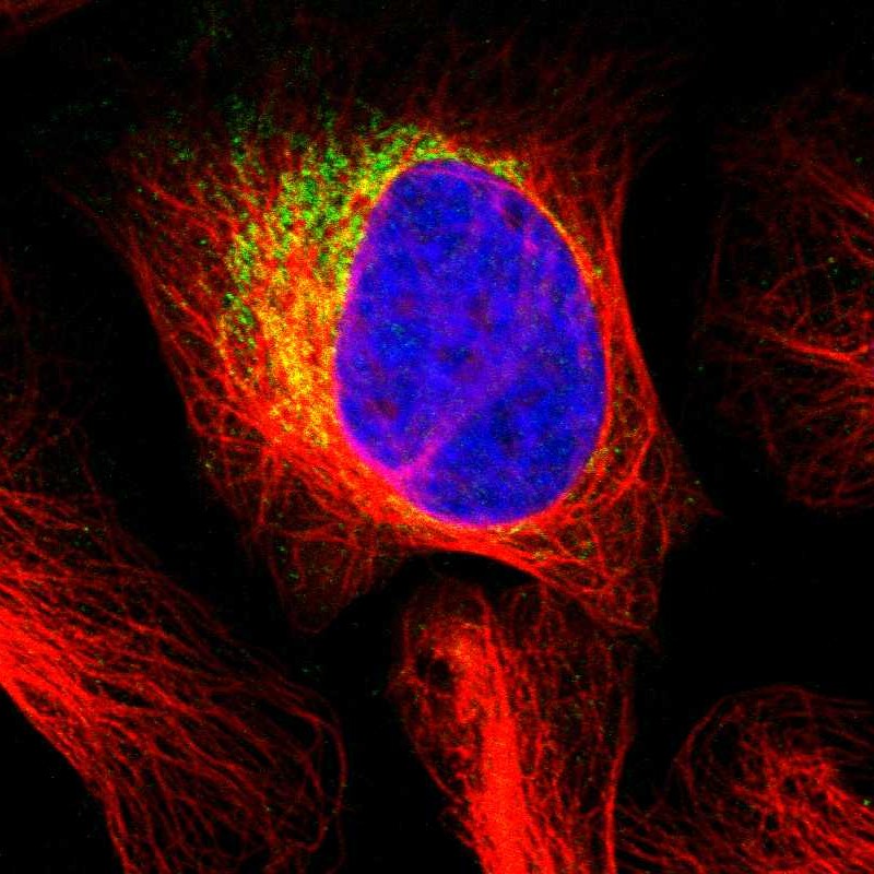
Immunofluorescent staining of human cell line U-2 OS shows localization to mitochondria.  

Antibody dilution

1:4  

Literature conformity

The subcellular location is supported by literature.  

IMMUNOHISTOCHEMISTRY


Antibody HPA016480


Standard validation

Approved  


Figure description

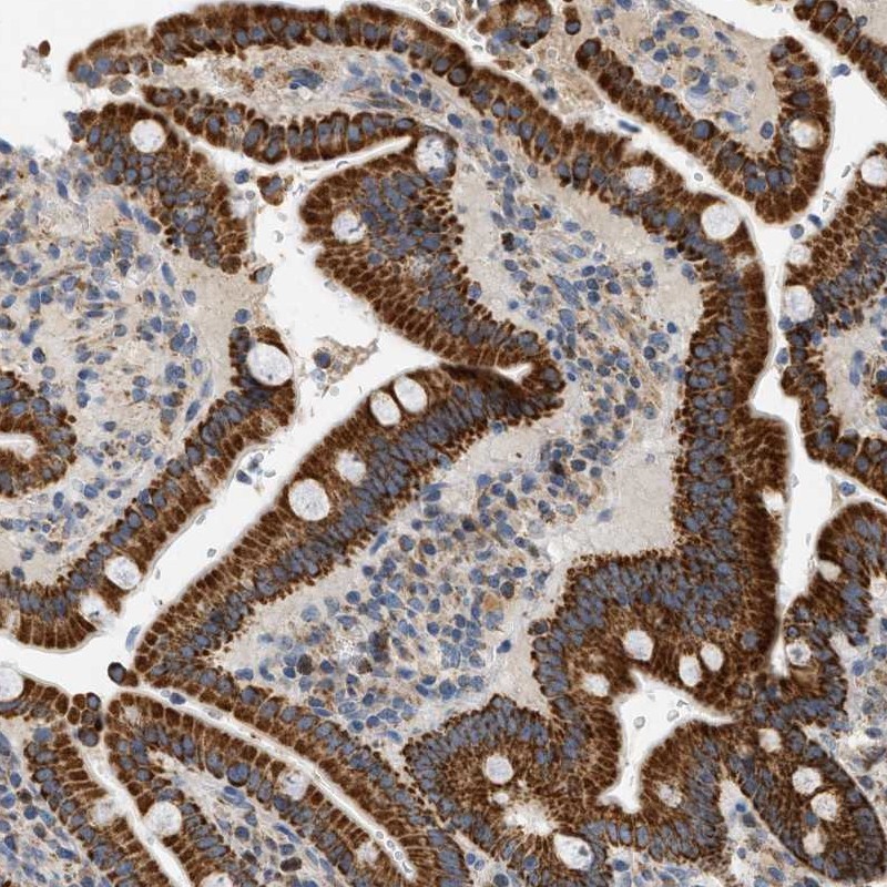
Immunohistochemical staining of human duodenum shows strong cytoplasmic positivity in glandular cells.  

Expression

RNA: detected in 37 tissues
Protein: detected in 79 cell types
 

Retrieval

HIER pH6  

Antibody dilution

1:7  

Literature conformity

Partly consistent with extensive gene/protein characterization data.  

RNA consistency

Mainly not consistent with RNA expression data.  

WESTERN BLOT


Antibody HPA016480


Standard validation

Supported

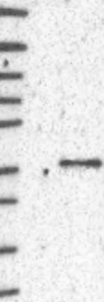
Single band corresponding to the predicted size in kDa (+/-20%).


Figure description

Lane 1: Marker [kDa] 250, 130, 95, 72, 55, 36, 28, 17, 10
Lane 2: Negative control (vector only transfected HEK293T lysate)
Lane 3: Over-expression Lysate (Co-expressed with a C-terminal myc-DDK tag (~3.1 kDa) in mammalian HEK293T cells, LY408629)
 

Target mass (kDa)

39.9, 37, 35.2  

Antibody dilution

1:125  

PROTEIN ARRAY


Antibody HPA016480


Standard validation

Supported

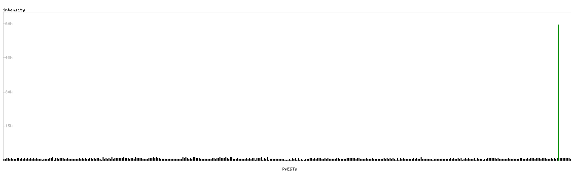
Pass with single peak corresponding to interaction only with its own antigen.


Figure description

Antibody specificity analysis with protein arrays. Predicted and matching interactions are shown in green.  

Antibody dilution

1:500  

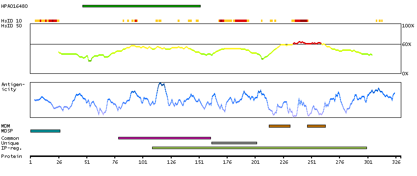
ANTIGEN INFORMATION


Antibody HPA016480


Antigen

Recombinant protein fragment  

Length (aa)

106  

Antigen sequence

HHRTVHQRIASWQNLGAVYCSTVVPSDDVTVVYQNGLPVISVRLPSRRER
CQFTLKPISDSVGVFLRQLQEEDRGIDRVAIYSPDGVRVAASTGIDLLLL
DDFKLV
 

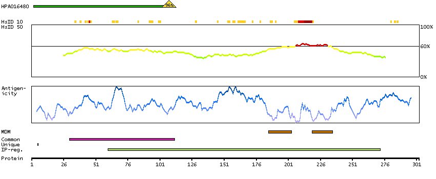
Matching transcripts

MCU-001 - ENSP00000349680 [100%]
MCU-002 - ENSP00000362144 [100%]
MCU-201 - ENSP00000440913 [96%]
 

Other gene match

 
ANTIGEN VIEW
MCU-001
MCU-002
MCU-201

RELEVANT PUBLICATIONS


Antibody HPA016480


1.

Integrative genomics identifies MCU as an essential component of the mitochondrial calcium uniporter
Baughman JM et al
Nature 2011;476(7360):341-5
 

2.

The physiological role of mitochondrial calcium revealed by mice lacking the mitochondrial calcium uniporter
Pan X et al
Nat Cell Biol 2013;15(12):1464-72
 

3.

Mitochondrial calcium uniporter Mcu controls excitotoxicity and is transcriptionally repressed by neuroprotective nuclear calcium signals
Qiu J et al
Nat Commun 2013;4:2034
 

4.

Loss-of-function mutations in MICU1 cause a brain and muscle disorder linked to primary alterations in mitochondrial calcium signaling
Logan CV et al
Nat Genet 2014;46(2):188-93
 

5.

Reconstitution of the mitochondrial calcium uniporter in yeast
Kovács-Bogdán E et al
Proc Natl Acad Sci U S A 2014;111(24):8985-90
 

6.

Chronic enrichment of hepatic endoplasmic reticulum-mitochondria contact leads to mitochondrial dysfunction in obesity
Arruda AP et al
Nat Med 2014;20(12):1427-35
Application: WB
 

7.

Quantitative proteomics of synaptic and nonsynaptic mitochondria: insights for synaptic mitochondrial vulnerability
Stauch KL et al
J Proteome Res 2014;13(5):2620-36
Application: WB
 

8.

Structure and function of the N-terminal domain of the human mitochondrial calcium uniporter
Lee Y et al
EMBO Rep 2015;16(10):1318-33
Application: WB
 

9.

The endoplasmic reticulum mitochondrial calcium cross talk is downregulated in malignant pleural mesothelioma cells and plays a critical role in apoptosis inhibition
Patergnani S et al
Oncotarget 2015;6(27):23427-44
Application: IHC, WB
 

10.

Essential role of mitochondrial Ca2+ uniporter in the generation of mitochondrial pH gradient and metabolism-secretion coupling in insulin-releasing cells
Quan X et al
J Biol Chem 2015;290(7):4086-96
Application: WB
 

11.

The mitochondrial calcium uniporter regulates breast cancer progression via HIF-1α
Tosatto A et al
EMBO Mol Med 2016;8(5):569-85
Application: WB


Contact

  • BLOG
  • PRESS ROOM
  • contact@proteinatlas.org

The Project

  • INTRODUCTION
  • ORGANIZATION
  • PUBLICATIONS
  • DOWNLOADABLE DATA

The Human Protein Atlas

  • ANTIBODY AVAILABILITY
  • LICENCE & CITATION
  • HELP & FAQ
The Human Protein Atlas project is funded
by the Knut & Alice Wallenberg foundation.