TISSUE
CELL
CANCER
ANTIBODY INFORMATION
Antibody HPA021560
Antibody HPA023295
Provider
Product name
Host species
Clonality
Purity
Other gene match
Released in version
References
VALIDATION SUMMARY
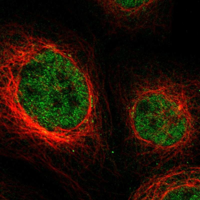
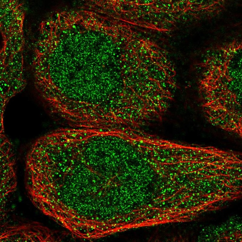
IMMUNOCYTOCHEMISTRY
Standard validation
Figure description
Antibody dilution
Literature conformity
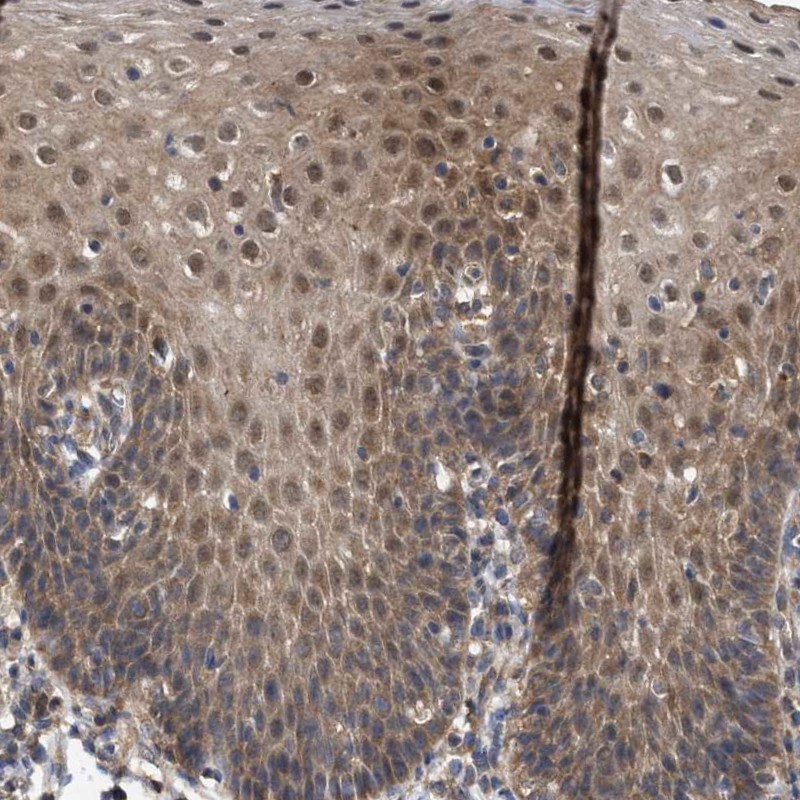
IMMUNOHISTOCHEMISTRY
Expression
Retrieval
RNA consistency
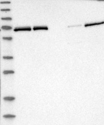
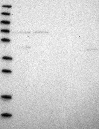
WESTERN BLOT
Target mass (kDa)
PROTEIN ARRAY
ANTIGEN INFORMATION
Antigen
Length (aa)
Antigen sequence
VRDECLLPCKDAPELGYAKESSSEQYVPDVFYKDVDKFGNEITQLARPLP
VEYLIIDITTTFPKDPVYTFSISQNPFPIENRDVLGETQDFHSLATYLSQ
NTSSVFLDTISDFHLLLFLVTNE
LEPFDEDYLNHLEPPVKHMSFHAYIRKLTGGADKGKFVALENISCKIKSG
CEGHLPWPNGICTKCQPSAITLNRQKYRHVDNIMFENHTVADRFLDFWRK
TGNQHFGYL
Matching transcripts
RELEVANT PUBLICATIONS
1.