We use cookies to enhance the usability of our website. If you continue, we'll assume that you are happy to receive all cookies. More information. Don't show this again.
Antibody staining overlaps with GFP tagged protein
Validated
Antibody staining overlaps with GFP tagged protein
Figure description
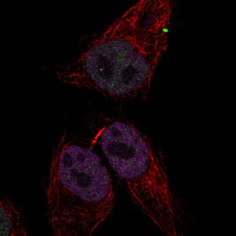
Immunofluorescent staining of transgenic HeLa cells show antibody staining in nucleus but excluded from the nucleoli and GFP expression in nucleus but excluded from the nucleoli.
Immunofluorescent staining of transgenic HeLa cells show antibody staining in nucleus but excluded from the nucleoli and GFP expression in nucleus but excluded from the nucleoli.
Antibody dilution
1:53
1:45
Formal validation: Independent
Validated
Antibody staining overlaps with antibody HPA062226.
Validated
Antibody staining overlaps with antibody HPA047549.
Standard validation
Supported
Supported
Figure description
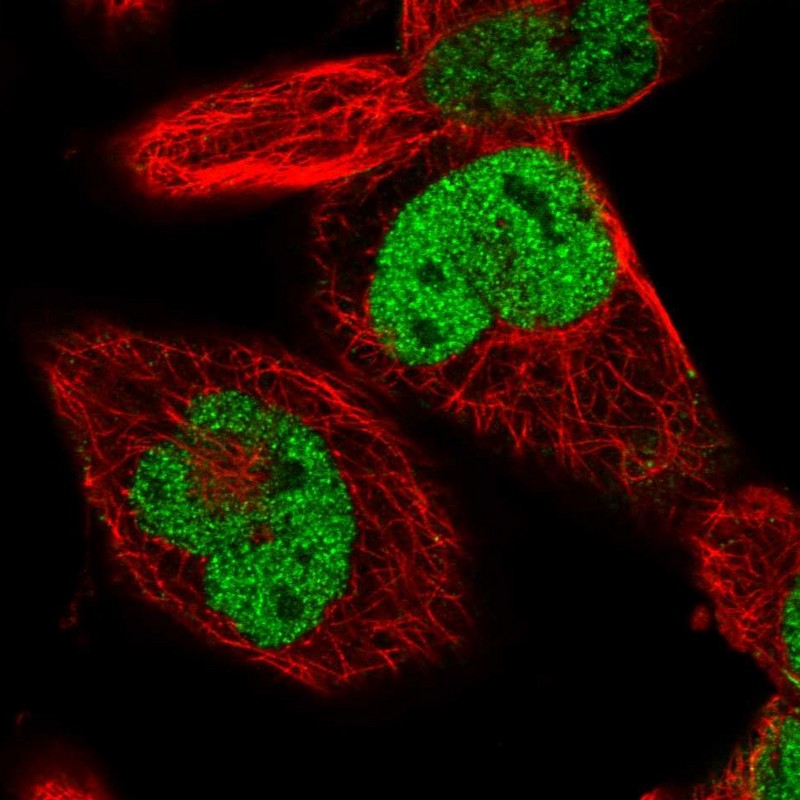
Immunofluorescent staining of human cell line HEK 293 shows localization to nucleoplasm.
Immunofluorescent staining of human cell line RH-30 shows localization to nucleoplasm.
Antibody dilution
1:53
1:45
Literature conformity
The subcellular location is supported by literature.
The subcellular location is supported by literature.
IMMUNOHISTOCHEMISTRY
Antibody HPA047549
Antibody HPA062226
Antibody CAB004254
Standard validation
Supported
Supported
Supported
Figure description
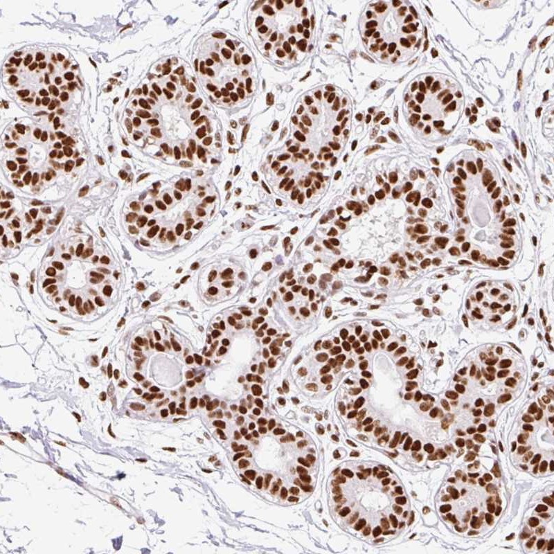
Immunohistochemical staining of human breast shows strong nuclear positivity in glandular cells and myoepithelial cells.
Immunohistochemical staining of human colon shows strong nuclear positivity in glandular cells.
Immunohistochemical staining of human colon shows strong nuclear positivity in glandular cells.
Expression
RNA: detected in 37 tissues Protein: detected in 78 cell types
RNA: detected in 37 tissues Protein: detected in 72 cell types
RNA: detected in 37 tissues Protein: detected in 75 cell types
Retrieval
HIER pH6
HIER pH6
HIER pH6
Antibody dilution
1:2500
1:75
1:3000
Literature conformity
Consistent with extensive gene/protein characterization data.
Consistent with extensive gene/protein characterization data.
Consistent with extensive gene/protein characterization data.
RNA consistency
Consistent with RNA expression data.
Consistent with RNA expression data.
Consistent with RNA expression data.
WESTERN BLOT
Antibody HPA047549
Antibody HPA062226
Antibody CAB004254
Formal validation: Genetic
Validated
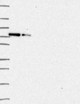
Downregulation visible in both siRNA lanes
Figure description
Lane 1: Marker [kDa] 250, 130, 95, 72, 55, 36, 28, 17, 10 Lane 2: siRNA 1 Lane 3: siRNA 2 Lane 4: Scrambled