We use cookies to enhance the usability of our website. If you continue, we'll assume that you are happy to receive all cookies. More information. Don't show this again.
Fields »
 

Field

Term
Gene name

Class

Subclass

Chromosome

External id

In atlas

Reliability

Reliability

Reliability

Validation

Method

Validation

Method

Validation

Method

Validation

Location

Searches

Tissue

Cell type

Expression

Tissue

Specificity

Cell line

Specificity

Tissue

Detectable

Cell line

Detectable

Category

Score

Score

Score

Antibodies

Column


ABOUT
HELP
BLOG
  • THE HUMAN PROTEOME
  • THE HUMAN TISSUES
  • THE HUMAN CELL
  • PROTEIN CLASSES
  • PROTEIN EVIDENCE
    • NEWS
    • BLOG
    • EVENTS
    • PRESS ROOM
    • LEARN
    • DICTIONARY
    • METHODS
    • CELL LINES
  • THE PROJECT
  • INTRODUCTION
  • ORGANIZATION
  • PUBLICATIONS
  • PUBLICATION DATA
  • ANTIBODY SUBMISSION
  • ANTIBODY AVAILABILITY
  • LINKS
  • CONTACT
  • TECHNICAL DATA
  • ANTIBODY VALIDATION
  • ASSAYS & ANNOTATION
  • DISCLAIMER
  • DOWNLOADABLE DATA
  • HELP & FAQ
  • LICENCE & CITATION
  • PRIVACY STATEMENT
  • RELEASE HISTORY
MYO1C
  • premium

    TISSUE

  • premium

    CELL

  • CANCER

ANTIBODY INFORMATION VALIDATION SUMMARY IMMUNOCYTOCHEMISTRY IMMUNOHISTOCHEMISTRY WESTERN BLOT PROTEIN ARRAY ANTIGEN INFORMATION RELEVANT PUBLICATIONS

ANTIBODY INFORMATION


Antibody HPA001768


Antibody HPA005924


Antibody CAB037023


Provider

Atlas Antibodies
Sigma-Aldrich
  Atlas Antibodies
Sigma-Aldrich
  SIGMA
 

Product name

HPA001768   HPA005924   M3567  

Host species

Rabbit   Rabbit   Rabbit  

Clonality

pAb   pAb   msAb  

Purity

Affinity purified using the PrEST-antigen as affinity ligand   Affinity purified using the PrEST-antigen as affinity ligand   Affinity  

Other gene match


Released in version

4   16   12  

References

3      

VALIDATION SUMMARY


Antibody HPA001768


Antibody HPA005924


Antibody CAB037023

 
Immunocytochemistry


Validated
   
Immunohistochemistry

Supported     Supported  
Western blot

Supported   Uncertain   Supported  
Protein array

Supported   Supported    

IMMUNOCYTOCHEMISTRY


Antibody HPA001768


Antibody HPA005924


Antibody CAB037023


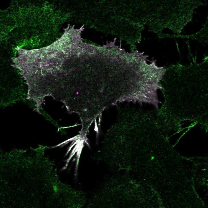
Formal validation: Tagged protein

 
Validated

Antibody staining overlaps with GFP tagged protein


Figure description

  Immunofluorescent staining of transgenic HeLa cells show antibody staining in plasma membrane and GFP expression in plasma membrane.    

Antibody dilution

  1:23    
 

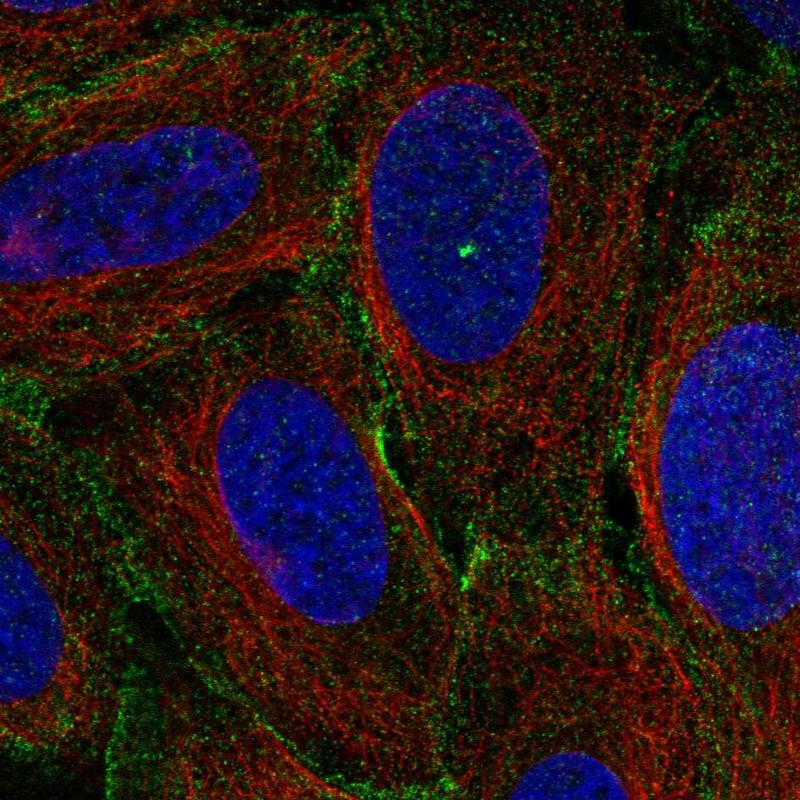
Standard validation

  Supported    


Figure description

  Immunofluorescent staining of human cell line U-2 OS shows localization to nuclear bodies & plasma membrane.    

Antibody dilution

  1:23    

Literature conformity

  The subcellular location is supported by literature.    

IMMUNOHISTOCHEMISTRY


Antibody HPA001768


Antibody HPA005924


Antibody CAB037023


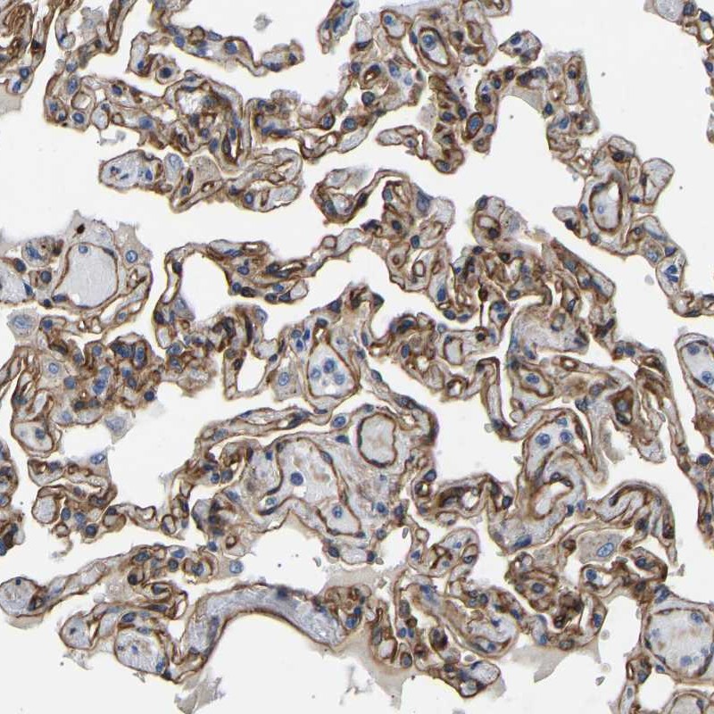
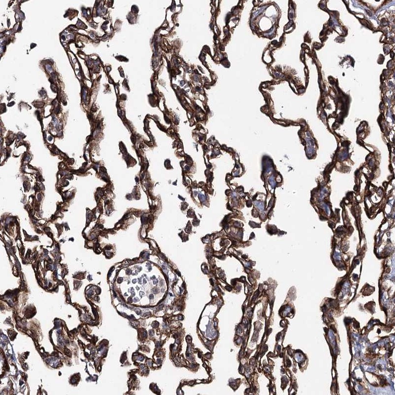
Standard validation

Supported     Supported  


Figure description

Immunohistochemical staining of human lung shows cytoplasmic and membranous positivity in pneumocytes.     Immunohistochemical staining of human lung shows cytoplasmic and membranous positivity in pneumocytes.  

Expression

RNA: detected in 37 tissues
Protein: detected in 72 cell types
    RNA: detected in 37 tissues
Protein: detected in 73 cell types
 

Retrieval

HIER pH6     HIER pH6  

Antibody dilution

1:75     1:350  

Literature conformity

Partly consistent with extensive gene/protein characterization data.     Partly consistent with extensive gene/protein characterization data.  

RNA consistency

Mainly consistent with RNA expression data.     Mainly consistent with RNA expression data.  

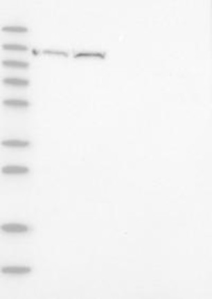
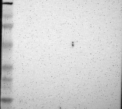
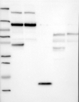
WESTERN BLOT


Antibody HPA001768


Antibody HPA005924


Antibody CAB037023


Standard validation

Supported

Analysis performed using a standard panel of samples.
Single band corresponding to the predicted size in kDa (+/-20%).
  Uncertain

Analysis performed using a standard panel of samples.
No bands detected.
  Supported

Analysis performed using a standard panel of samples.
Band of predicted size in kDa (+/-20%) with additional bands present.


Figure description

Lane 1: Marker [kDa] 230, 130, 95, 72, 56, 36, 28, 17, 11
Lane 2: RT4
Lane 3: U-251 MG
Lane 4: Human Plasma
Lane 5: Liver
Lane 6: Tonsil
  Lane 1: Marker [kDa] 220, 112, 84, 47, 32, 26, 16.8
Lane 2: RT4
Lane 3: U-251 MG
Lane 4: Human Plasma
Lane 5: Liver
Lane 6: Tonsil
  Lane 1: Marker [kDa] 250, 130, 100, 70, 55, 35, 25, 15, 10
Lane 2: RT4
Lane 3: U-251 MG
Lane 4: Human Plasma
Lane 5: Liver
Lane 6: Tonsil
 

Target mass (kDa)

121.7, 119.6, 119, 117.9   121.7, 119.6, 119, 117.9   121.7, 119.6, 119, 117.9, 30.7, 19.5, 19.2, 18.7, 15.9, 15.4  

Antibody dilution

1:250   1:250   1:500  

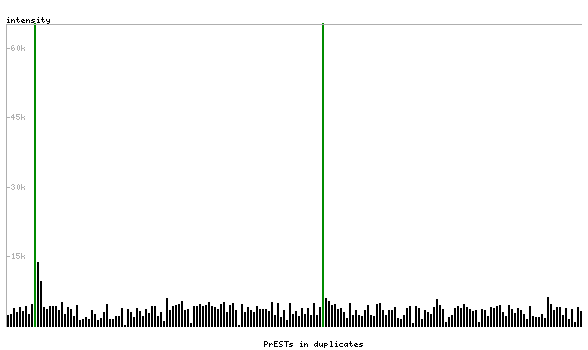
PROTEIN ARRAY


Antibody HPA001768


Antibody HPA005924


Antibody CAB037023


Standard validation

Supported

Pass with single peak corresponding to interaction only with its own antigen.
  Supported

Pass with single peak corresponding to interaction only with its own antigen.


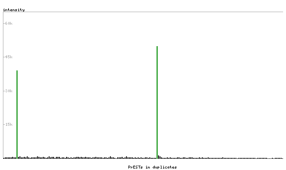
Figure description

Antibody specificity analysis with protein arrays. Predicted and matching interactions are shown in green.   Antibody specificity analysis with protein arrays. Predicted and matching interactions are shown in green.    

Antibody dilution

1:3000   1:3000    

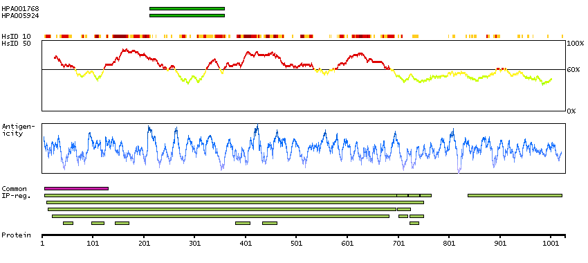
ANTIGEN INFORMATION


Antibody HPA001768


Antibody HPA005924


Antibody CAB037023


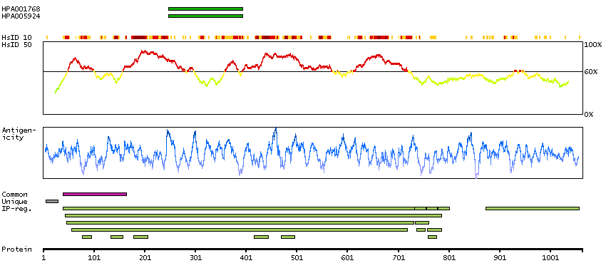
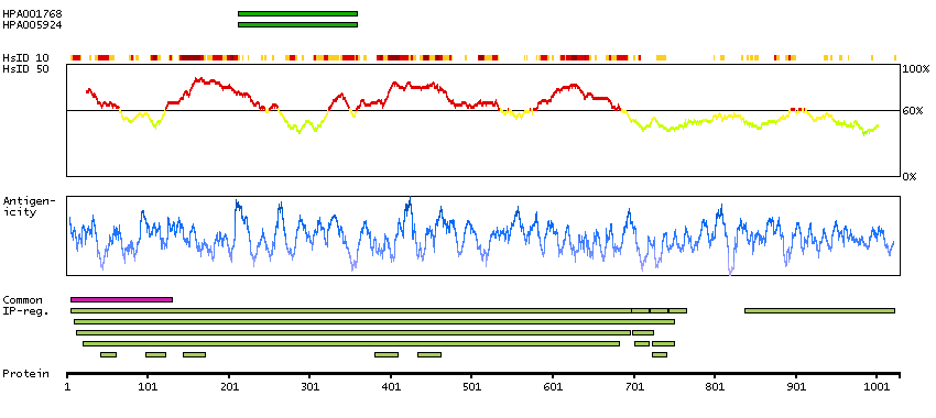
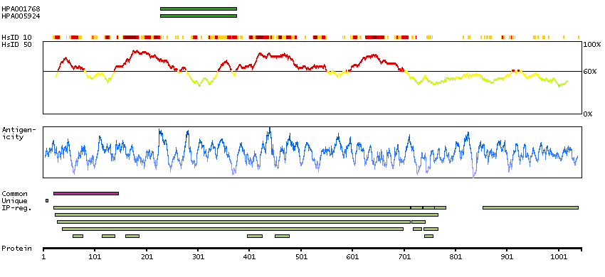
Antigen

Recombinant protein fragment   Recombinant protein fragment   Synthetic peptide  

Length (aa)

148   148    

Antigen sequence

GEEETLRRLGLERNPQSYLYLVKGQCAKVSSINDKSDWKVVRKALTVIDF
TEDEVEDLLSIVASVLHLGNIHFAANEESNAQVTTENQLKYLTRLLSVEG
STLREALTHRKIIAKGEELLSPLNLEQAAYARDALAKAVYSRTFTWLV
 
GEEETLRRLGLERNPQSYLYLVKGQCAKVSSINDKSDWKVVRKALTVIDF
TEDEVEDLLSIVASVLHLGNIHFAANEESNAQVTTENQLKYLTRLLSVEG
STLREALTHRKIIAKGEELLSPLNLEQAAYARDALAKAVYSRTFTWLV
   

Matching transcripts

MYO1C-001 - ENSP00000352834 [100%]
MYO1C-002 - ENSP00000459174 [100%]
MYO1C-003 - ENSP00000437685 [100%]
MYO1C-010 - ENSP00000412197 [100%]
MYO1C-201 - ENSP00000354283 [100%]
  MYO1C-001 - ENSP00000352834 [100%]
MYO1C-002 - ENSP00000459174 [100%]
MYO1C-003 - ENSP00000437685 [100%]
MYO1C-010 - ENSP00000412197 [100%]
MYO1C-201 - ENSP00000354283 [100%]
   

Other gene match

     
ANTIGEN VIEW
MYO1C-001
MYO1C-002
MYO1C-003
MYO1C-010
MYO1C-201

RELEVANT PUBLICATIONS


Antibody HPA001768


Antibody HPA005924


Antibody CAB037023


1.

The myosin motor Myo1c is required for VEGFR2 delivery to the cell surface and for angiogenic signaling
Tiwari A et al
Am J Physiol Heart Circ Physiol 2013;304(5):H687-96
     

2.

Analysis of an independent tumor suppressor locus telomeric to Tp53 suggested Inpp5k and Myo1c as novel tumor suppressor gene candidates in this region
Hedberg Oldfors C et al
BMC Genet 2015;16:80
Application: WB
     

3.

Loss of functional MYO1C/myosin 1c, a motor protein involved in lipid raft trafficking, disrupts autophagosome-lysosome fusion
Brandstaetter H et al
Autophagy 2014;10(12):2310-23
Application: WB


Contact

  • BLOG
  • PRESS ROOM
  • contact@proteinatlas.org

The Project

  • INTRODUCTION
  • ORGANIZATION
  • PUBLICATIONS
  • DOWNLOADABLE DATA

The Human Protein Atlas

  • ANTIBODY AVAILABILITY
  • LICENCE & CITATION
  • HELP & FAQ
The Human Protein Atlas project is funded
by the Knut & Alice Wallenberg foundation.