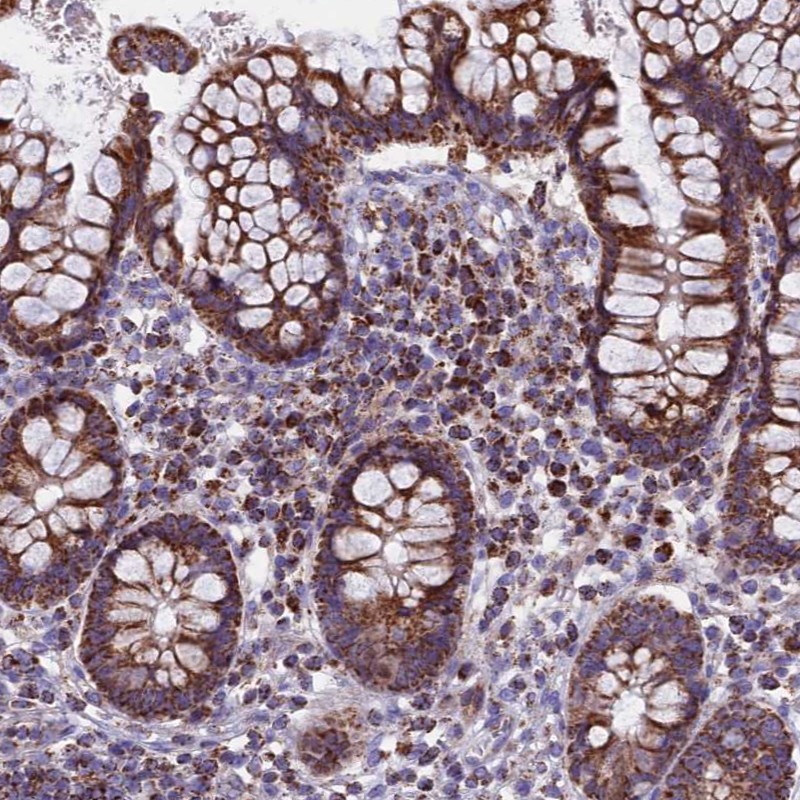
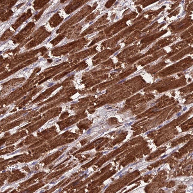
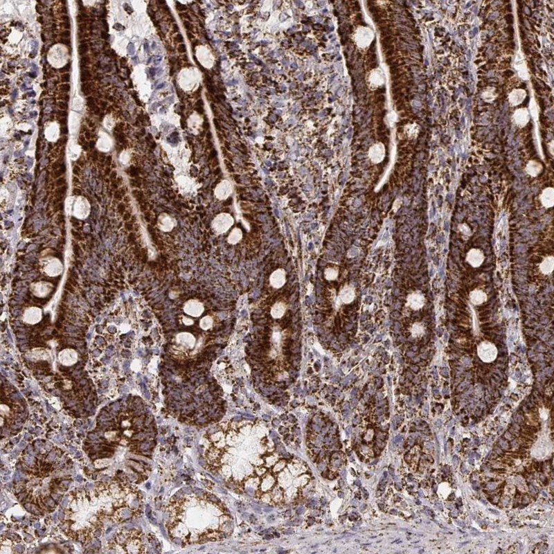
TISSUE
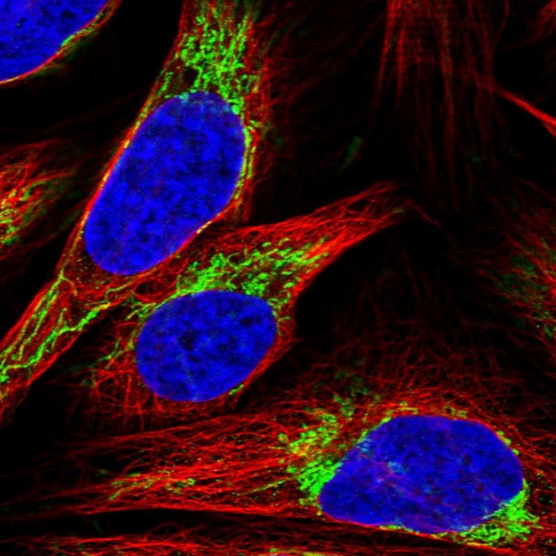
CELL
CANCER
ANTIBODY INFORMATION
Antibody HPA038460
Antibody HPA038461
Antibody CAB075750
Antibody CAB075751
Antibody CAB075752
Antibody CAB075753
Antibody CAB075754
Provider
Product name
Host species
Clonality
Purity
Other gene match
Released in version
References
VALIDATION SUMMARY
IMMUNOCYTOCHEMISTRY
Formal validation: Genetic
Relative fluorescence intensity (RFI)
Figure description
Antibody dilution
Formal validation: Independent
Standard validation
Literature conformity
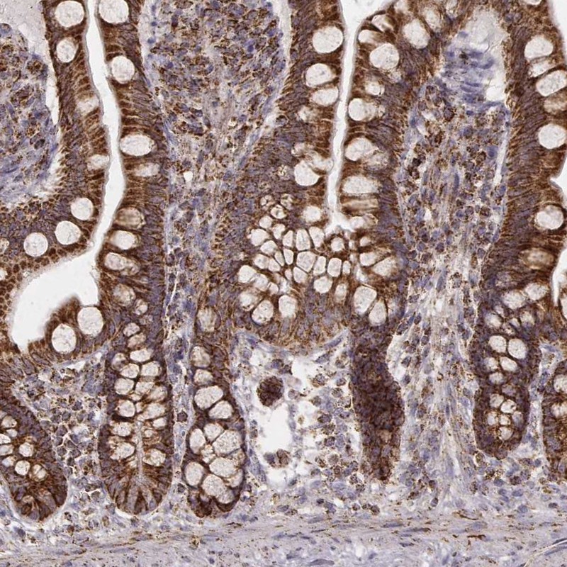
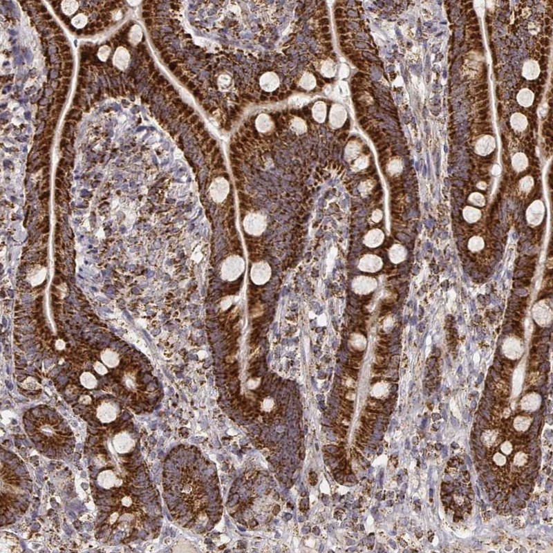
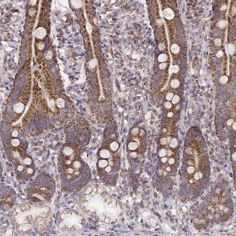
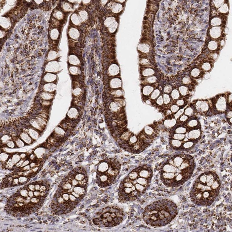
IMMUNOHISTOCHEMISTRY
Expression
Retrieval
RNA consistency
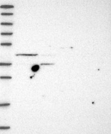
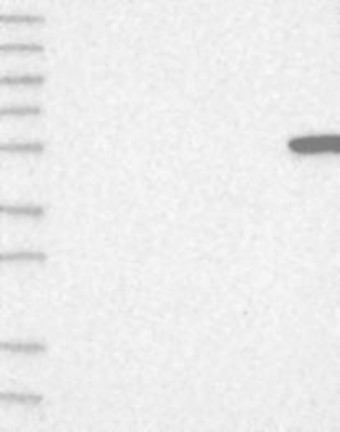
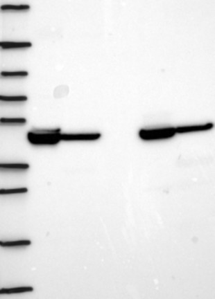
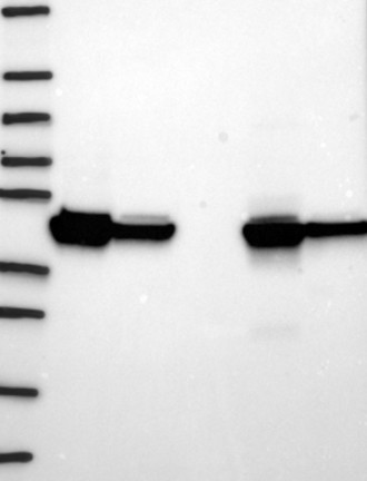
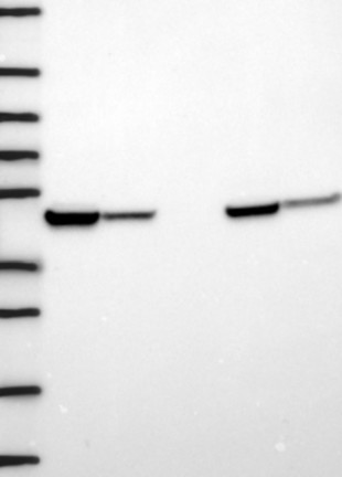
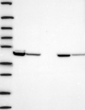
WESTERN BLOT
Target mass (kDa)
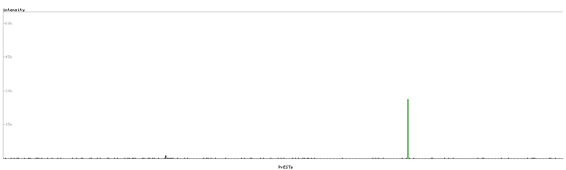
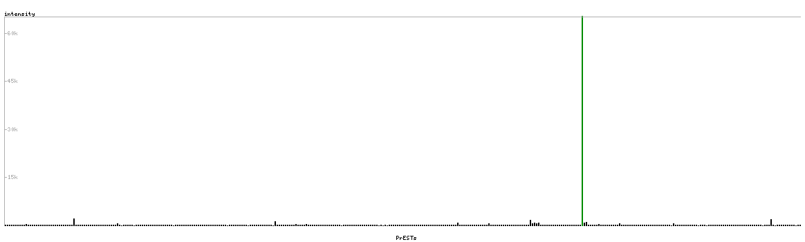
PROTEIN ARRAY
ANTIGEN INFORMATION
Antigen
Length (aa)
Antigen sequence
LDWSHNFTNMLGYTDHQFTELTRLYLTIHSDHEGGNVSAHTSHLVGSALS
DPYLSFAAAMNGLAGPLHGLANQEVLVWLTQLQKEVGKDVS
ADLIPKEQARIKTFRQQHGKTVVGQITVDMMYGGMRGMKGLVYETSVLDP
DEGIRFRGFSIPECQKLLPKAKGGEEPLPEGLFWLLVTGHIPTEEQVSWL
Matching transcripts