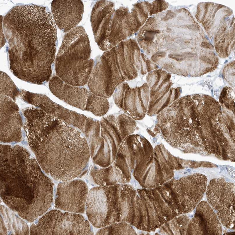
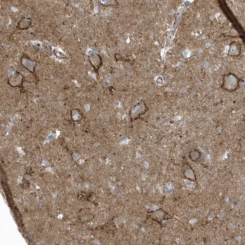
TISSUE
CELL
CANCER
ANTIBODY INFORMATION
Antibody HPA035325
Antibody HPA035326
Provider
Product name
Host species
Clonality
Purity
Other gene match
Released in version
References
VALIDATION SUMMARY
IMMUNOHISTOCHEMISTRY
Standard validation
Figure description
Expression
Retrieval
Antibody dilution
Literature conformity
RNA consistency
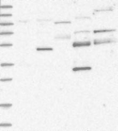
WESTERN BLOT
Target mass (kDa)
PROTEIN ARRAY
ANTIGEN INFORMATION
Antigen
Length (aa)
Antigen sequence
SLGDIVPSSRKSTPPSSAIDIDATGLDAEENDIPANHRSPKPSANSVTSP
RDSAYVEPKEDYSHDHVDHYASHRDHNHRDETHGSSDHRHRESRHRSRDV
DREQDHNECNKQRSRHKSKDRYCEKDGEVI
Matching transcripts