TISSUE
CELL
CANCER
ANTIBODY INFORMATION
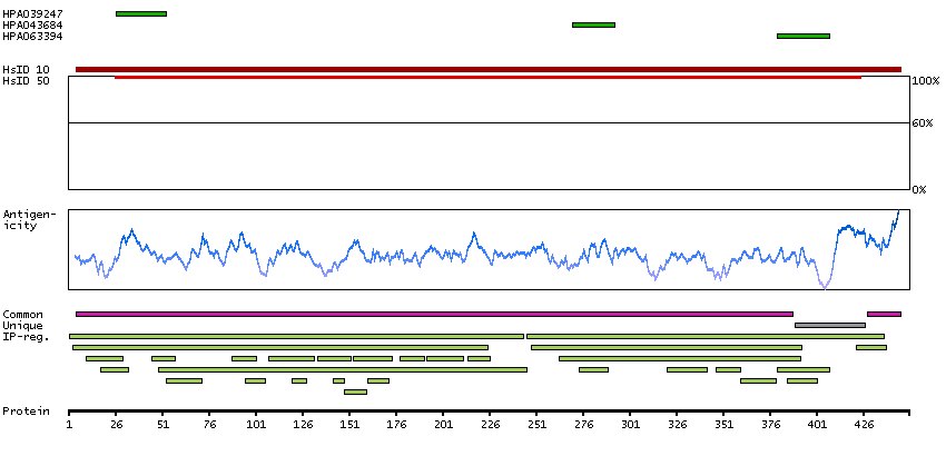
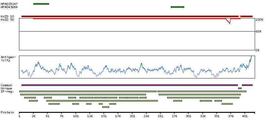
Antibody HPA039247
Antibody HPA043684
Antibody HPA063394
Provider
Product name
Host species
Clonality
Purity
Other gene match
Released in version
References
VALIDATION SUMMARY
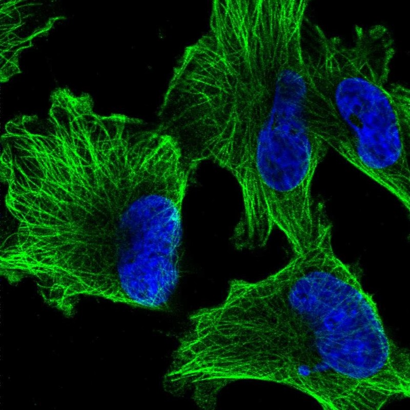
IMMUNOCYTOCHEMISTRY
Standard validation
Figure description
Antibody dilution
Literature conformity
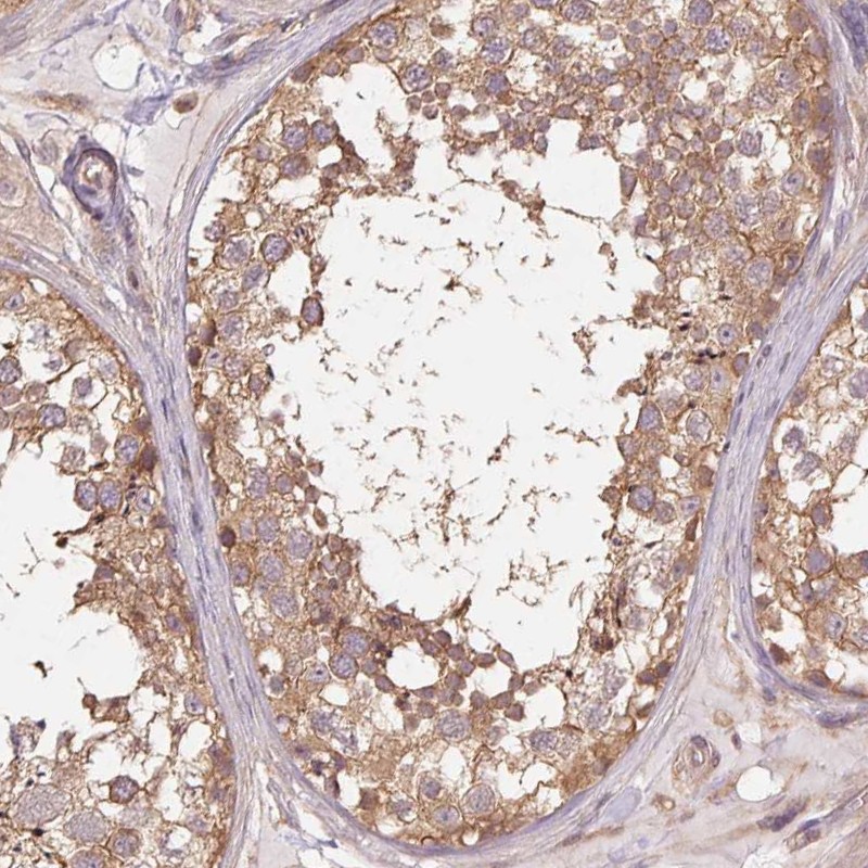
IMMUNOHISTOCHEMISTRY
Expression
Retrieval
RNA consistency
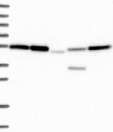
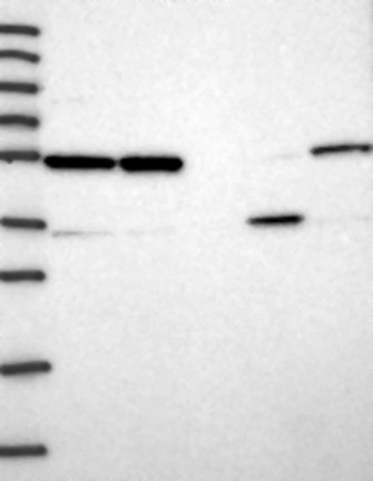
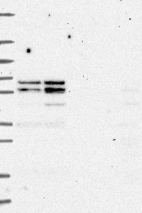
WESTERN BLOT
Target mass (kDa)
PROTEIN ARRAY
ANTIGEN INFORMATION
Antigen
Length (aa)
Antigen sequence
LEHGIQPDGQMPSDKTIGGGDDSFNTFF
ATYAPVISAEKAYHEQLSVAEITN
NTTAIAEAWARLDHKFDLMYAKRAFVHWY
Matching transcripts